. 2016 Mar;13(3):2467-75.
doi: 10.3892/mmr.2016.4859. Epub 2016 Feb 4.

Association of the hERG mutation with long-QT syndrome type 2, syncope and epilepsy

Affiliations

Association of the hERG mutation with long-QT syndrome type 2, syncope and epilepsy

Guoliang Li et al. Mol Med Rep. 2016 Mar.

Abstract

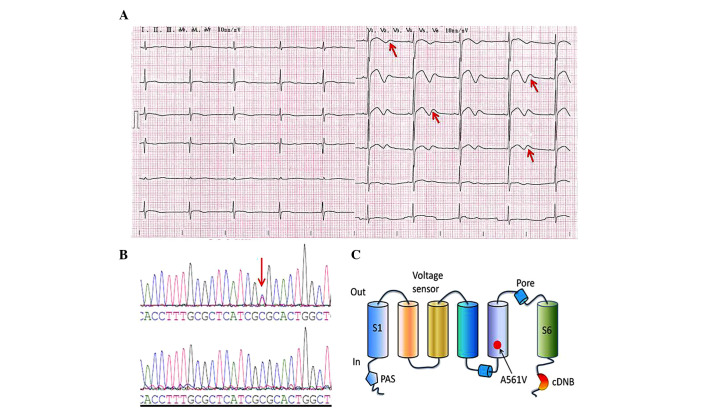
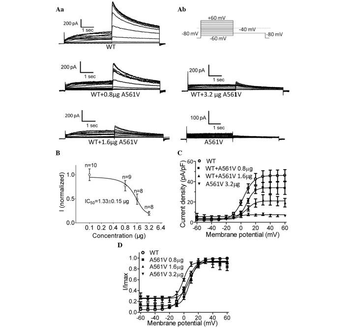
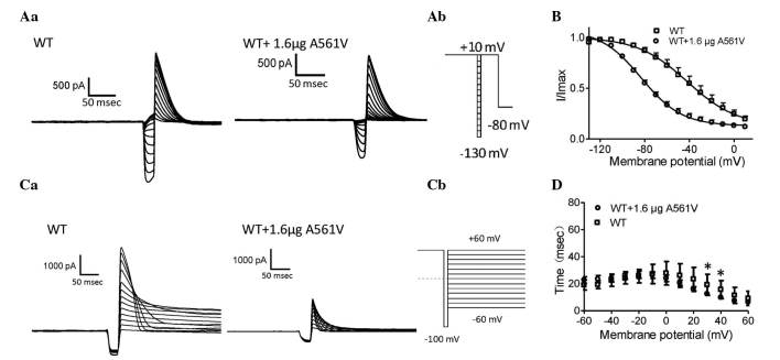
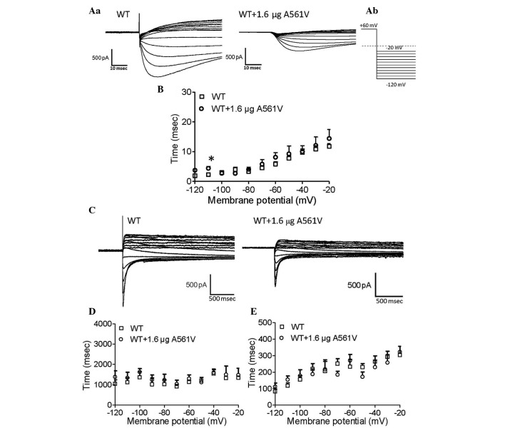
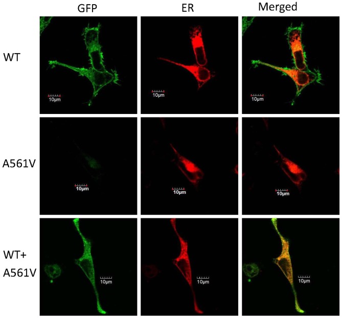
Mutations in the human ether‑à‑go‑go‑related gene (hERG) are responsible for long‑QT syndrome (LQTS) type 2 (LQT2). In the present study, a heterozygous missense mutation (A561V) linked to LQT2, syncope and epilepsy was identified in the S5/pore region of the hERG protein. The mutation, A561V, was prepared and subcloned into hERG‑pcDNA3.0. Mutant plasmids were co‑transfected into HEK‑293 cells, which stably express wild‑type (WT) hERG, in order to mimic a heterozygous genotype, and the whole‑cell current was recorded using a patch‑clamp technique. Confocal microscopy was performed to evaluate the membrane distribution of the hERG channel protein using a green fluorescent protein tagged to the N‑terminus of hERG. A561V‑hERG decreased the amplitude of the WT‑hERG currents in a concentration‑dependent manner. In addition, A561V‑hERG resulted in alterations to activation, inactivation and recovery from inactivation in the hERG protein channels. Further evaluation of hERG membrane localization indicated that the A561V‑hERG mutant protein was unable to travel to the plasma membrane, which resulted in a trafficking‑deficient WT‑hERG protein. In conclusion, A561V‑hERG exerts a potent dominant‑negative effect on WT‑hERG channels, resulting in decreased hERG currents and impairment of hERG membrane localization. This may partially elucidate the clinical manifestations of LQTS patients who carry the A561V mutation.


Figures

Figure 1
Figure 1
ECG and mutation analysis. (A) ECG of the LQTS patient. The 12-lead ECG revealed a prolonged heart rate-corrected QT interval and biphasic T waves (indicated by the red arrows). (B) DNA sequencing chromatogram demonstrating a heterozygous point mutation, 1682C→T in the hERG gene of the LQTS patient (upper DNA sequencing chromatogram) and DNA sequence analysis in a healthy individual (lower DNA sequencing chromatogram); the red arrow indicates the mutation site. (C) This mutation leads to the substitution of the amino acid, alanine (A) by valine (V) in the S5/pore region of the hERG protein (A561V). ECG, electrocardiogram; LQTS, long-QT syndrome; hERG, human ether-à-go-go-related gene; cDNB, cyclic-nucleotide-binding domain.
Figure 2
Figure 2
A561V mutation dose-dependently suppressed the currents in HEK-293 cells stably expressing WT-hERG. (Aa) Representative whole-cell current traces obtained from cells stably expressing WT-hERG with and without varying quantities of A561V-hERG plasmid (0.8, 1.6 and 3.2 µg), as well as the currents in cells exhibiting the A561V mutation. (Ab) A portion of the voltage-clamp protocol. (B) Dose-dependent inhibition of hERG currents by the A561V mutation. (C) I–V associations of the tail currents obtained from WT-hERG cells and A561V-hERG plasmid-transfected WT-hERG cells. (D) The I–V curves were normalized to a Boltzmann function. WT, wild-type; hERG, human ether-à-go-go-related gene; I, current; V, voltage.
Figure 3
Figure 3
Steady-state inactivation and time courses of inactivation analysis. (Aa) Representative steady-state inactivation current traces. (Ab) A portion of the voltage-clamp protocol. (B) Normalized steady-state inactivation curves for WT-hERG and 1.6-µg A561V-transfected WT-hERG cells, respectively. (Ca) Representative current traces of the time courses of inactivation and inactivation time constant values (τ) for WT-hERG and 1.6-µg A561V-transfected WT-hERG cells. (Cb) A portion of the voltage-clamp protocol. (D) Normalized inactivation time constant, τ, in cells expressing WT-hERG and 1.6-µg A561V-transfected WT-hERG cells. τ was measured by fitting the inactivating currents, during test pulses at each potential, with a single exponential function. *P<0.05, WT vs. A561V-transfected WT group. WT, wild-type; hERG, human ether-à-go-go-related gene; I, current; V, voltage.
Figure 4
Figure 4
Kinetics of recovery from inactivation and deactivation analysis. (Aa) Representative current traces showing recovery from inactivation were obtained from WT-hERG and 1.6-µg A561V-transfected WT-hERG cells. (Ab) A portion of the voltage-clamp protocol. (B) Time constant (τ) values of recovery from inactivation for WT-hERG and 1.6-µg A561V-transfected WT-hERG cells. (C) Representative current recordings of deactivation in WT-hERG and 1.6-µg A561V-transfected WT-hERG cells. (D) Fast and (E) slow deactivation components of deactivation time constants (τ) as a function of test potential in WT-hERG and 1.6-µg A561V-transfected WT-hERG cells. *P<0.05, WT vs. A561V-transfected WT group. WT, wild-type; hERG, human ether-à-go-go-related gene; I, current; V, voltage.
Figure 5
Figure 5
Representative images of subcellular localization of the hERG protein expressed in HEK-293 cells. Top panel, HEK-293 cells expressing pEGFP-C2-WT; middle panel, HEK-293 cells expressing pEGFP-C2-A561V; and bottom panel, HEK-293 cells expressing pEGFP-C2-WT and pEGFP-C2-A561V. Left column, the hERG protein tagged with green fluorescence (green); middle column, HEK-293 cells transfected with pDsRed2-ER (red); and right column, merge of the two. Scale bar, 10 µm. GFP, green fluorescent protein; ER, endoplasmic reticulum, WT, wild-type.

References

    1. Li G, Cheng G, Wu J, Zhou X, Liu P, Sun C. Drug-induced long QT syndrome in women. Adv Ther. 2013;30:793–802. doi: 10.1007/s12325-013-0056-x. - DOI - PubMed
    1. Liu L, Hayashi K, Kaneda T, Ino H, Fujino N, Uchiyama K, Konno T, Tsuda T, Kawashiri MA, Ueda K, et al. A novel mutation in the transmembrane nonpore region of the KCNH2 gene causes severe clinical manifestations of long QT syndrome. Heart Rhythm. 2013;10:61–67. doi: 10.1016/j.hrthm.2012.09.053. - DOI - PubMed
    1. Nakano Y, Shimizu W. Genetics of long-QT syndrome. J Hum Genet. 2015 Jun 25; Epub ahead of print. - PubMed
    1. Zhang A, Sun C, Zhang L, Lv Y, Xue X, Li G, Cui C, Yan GX. L539 fs/47, a truncated mutation of human ether-a-go-go-related gene (hERG), decreases hERG ion channel currents in HEK 293 cells. Clin Exp Pharmacol Physiol. 2013;40:28–36. doi: 10.1111/1440-1681.12028. - DOI - PubMed
    1. Yang HT, Sun CF, Cui CC, Xue XL, Zhang AF, Li HB, Wang DQ, Shu J. HERG-F463L potassium channels linked to long QT syndrome reduce I(Kr) current by a trafficking-deficient mechanism. Clin Exp Pharmacol Physiol. 2009;36:822–827. doi: 10.1111/j.1440-1681.2009.05150.x. - DOI - PubMed

Publication types

Substances

Supplementary concepts