

Classical scrapie prions are associated with peripheral blood monocytes and T-lymphocytes from naturally infected sheep
- PMID: 26847623
- PMCID: PMC4743119
- DOI: 10.1186/s12917-016-0651-6
Classical scrapie prions are associated with peripheral blood monocytes and T-lymphocytes from naturally infected sheep
Abstract
Background: Classical scrapie is a transmissible spongiform encephalopathy (TSE) that affects sheep and goats. Our previous bioassay studies in lambs revealed that scrapie prions could be detected in association with peripheral blood monocular cells (PBMC), B lymphocytes and platelet-rich plasma fractions. In the present study, bioassay in lambs was again used to determine if scrapie prions are associated with the other two subsets of PBMC, monocytes and T lymphocytes.
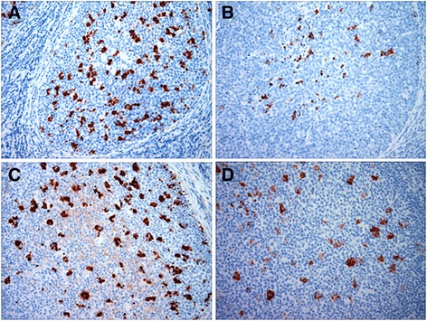
Results: PBMC, monocytes and T lymphocytes were isolated from two preclinically affected VRQ/VRQ sheep naturally infected with classical ovine scrapie and intravenously transfused into VRQ/VRQ lambs post-weaning. As determined using standard immunohistochemistry for scrapie, abnormal isoforms of prion protein were detected in lymphoid tissues of lambs inoculated with PBMC (4/4 recipient lambs), monocytes (2/5) and T lymphocytes (1/4). Prion protein misfolding activity was detected by serial protein misfolding cyclic amplification (sPMCA) in PBMC from monocyte and T lymphocyte recipient sheep in agreement with antemortem rectal biopsy results, but such prion protein misfolding activity was not detected from other recipients.
Conclusions: These findings show that scrapie prions are associated with monocytes and T lymphocytes circulating in the peripheral blood of sheep naturally infected with classical scrapie. Combined with our previous findings, we can now conclude that all three major subsets of PBMC can harbor prions during preclinical disease and thus, present logical targets for development of a sensitive assay to detect scrapie prions. In this regard, we have also demonstrated that sPMCA can be used to detect scrapie prions associated with PBMC.
Figures
Similar articles
-
Classical scrapie prions in ovine blood are associated with B lymphocytes and platelet-rich plasma.BMC Vet Res. 2011 Nov 23;7:75. doi: 10.1186/1746-6148-7-75. BMC Vet Res. 2011. PMID: 22112371 Free PMC article.
-
Cellular prion protein is expressed on peripheral blood mononuclear cells but not platelets of normal and scrapie-infected sheep.Haematologica. 2001 Feb;86(2):146-53. Haematologica. 2001. PMID: 11224483
-
Classical natural ovine scrapie prions detected in practical volumes of blood by lamb and transgenic mouse bioassays.J Vet Sci. 2015;16(2):179-86. doi: 10.4142/jvs.2015.16.2.179. Epub 2014 Dec 24. J Vet Sci. 2015. PMID: 25549221 Free PMC article.
-
Prion encephalopathies of animals and humans.Dev Biol Stand. 1993;80:31-44. Dev Biol Stand. 1993. PMID: 8270114 Review.
-
[Biology of non-conventional transmissible agents or prions].Rev Neurol (Paris). 1998 Feb;154(2):142-51. Rev Neurol (Paris). 1998. PMID: 9773035 Review. French.
Cited by
-
Modulating the Structure and Function of an Aminoacyl-tRNA Synthetase Cofactor by Biotinylation.J Biol Chem. 2016 Aug 12;291(33):17102-11. doi: 10.1074/jbc.M116.734343. Epub 2016 Jun 21. J Biol Chem. 2016. PMID: 27330079 Free PMC article.
-
Polymorphism of prion protein gene (PRNP) in Nigerian sheep.Prion. 2023 Dec;17(1):44-54. doi: 10.1080/19336896.2023.2186767. Prion. 2023. PMID: 36892181 Free PMC article.
-
Proteomic analysis revealed T cell hyporesponsiveness induced by Haemonchus contortus excretory and secretory proteins.Vet Res. 2020 May 13;51(1):65. doi: 10.1186/s13567-020-00790-0. Vet Res. 2020. PMID: 32404195 Free PMC article.
-
Detection of Pathognomonic Biomarker PrPSc and the Contribution of Cell Free-Amplification Techniques to the Diagnosis of Prion Diseases.Biomolecules. 2020 Mar 19;10(3):469. doi: 10.3390/biom10030469. Biomolecules. 2020. PMID: 32204429 Free PMC article. Review.
-
Cerebrospinal Fluid and Plasma Small Extracellular Vesicles and miRNAs as Biomarkers for Prion Diseases.Int J Mol Sci. 2021 Jun 25;22(13):6822. doi: 10.3390/ijms22136822. Int J Mol Sci. 2021. PMID: 34201940 Free PMC article.
References
-
- Andreoletti O, Berthon P, Marc D, Sarradin P, Grosclaude J, van Keulen L, et al. Early accumulation of PrP(Sc) in gut-associated lymphoid and nervous tissues of susceptible sheep from a Romanov flock with natural scrapie. J Gen Virol. 2000;81(Pt 12):3115–3126. doi: 10.1099/0022-1317-81-12-3115. - DOI - PubMed
Publication types
MeSH terms
Substances
LinkOut - more resources
Full Text Sources
Other Literature Sources
Research Materials
