. 2016 Apr;36(4):598-607.
doi: 10.1161/ATVBAHA.115.306931. Epub 2016 Feb 4.

Macrophage IKKα Deficiency Suppresses Akt Phosphorylation, Reduces Cell Survival, and Decreases Early Atherosclerosis

Affiliations

Macrophage IKKα Deficiency Suppresses Akt Phosphorylation, Reduces Cell Survival, and Decreases Early Atherosclerosis

Vladimir R Babaev et al. Arterioscler Thromb Vasc Biol. 2016 Apr.

Abstract

Objective: The IκB kinase (IKK) is an enzyme complex that initiates the nuclear factor κB transcription factor cascade, which is important in regulating multiple cellular responses. IKKα is directly associated with 2 major prosurvival pathways, PI3K/Akt and nuclear factor κB, but its role in cell survival is not clear. Macrophages play critical roles in the pathogenesis of atherosclerosis, yet the impact of IKKα signaling on macrophage survival and atherogenesis remains unclear.

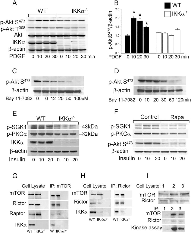
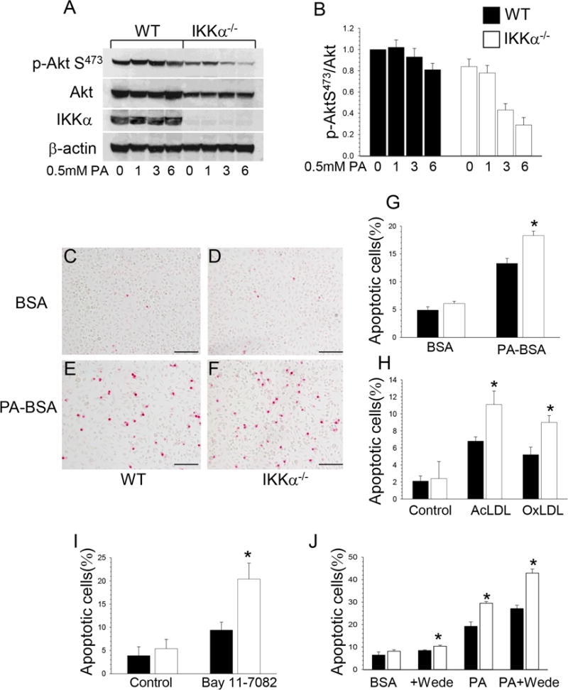
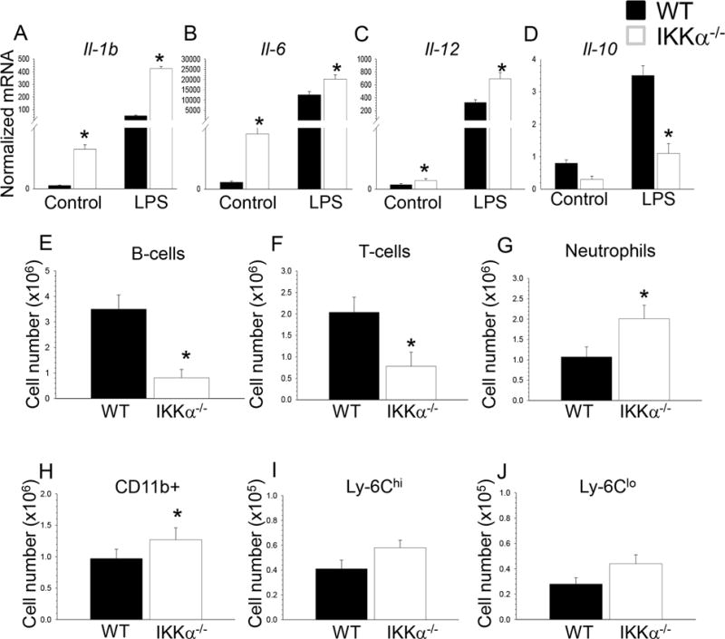
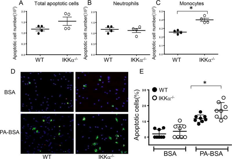
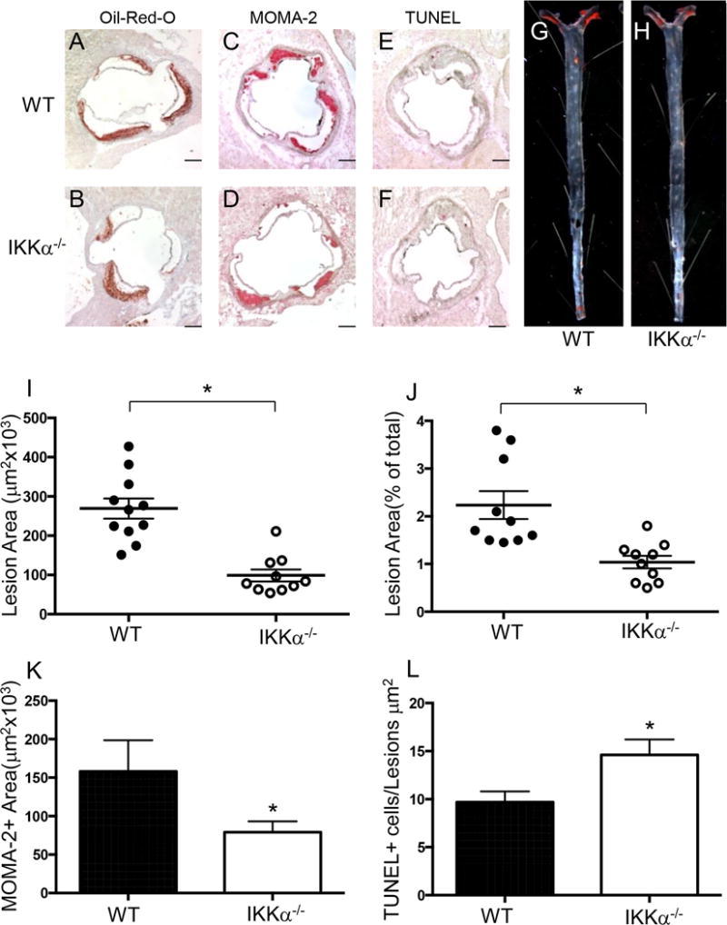
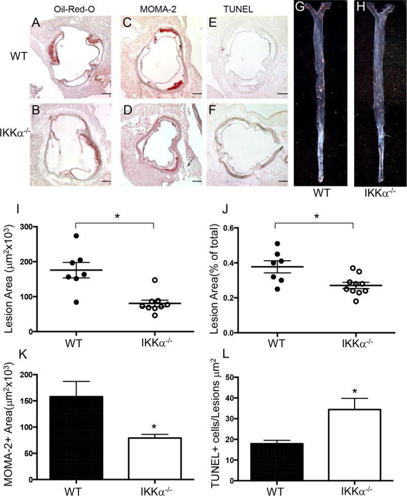
Approach and results: Here, we demonstrate that genetic IKKα deficiency, as well as pharmacological inhibition of IKK, in mouse macrophages significantly reduces Akt S(473) phosphorylation, which is accompanied by suppression of mTOR complex 2 signaling. Moreover, IKKα null macrophages treated with lipotoxic palmitic acid exhibited early exhaustion of Akt signaling compared with wild-type cells. This was accompanied by a dramatic decrease in the resistance of IKKα(-/-) monocytes and macrophages to different proapoptotic stimuli compared with wild-type cells. In vivo, IKKα deficiency increased macrophage apoptosis in atherosclerotic lesions and decreased early atherosclerosis in both female and male low-density lipoprotein receptor (LDLR)(-/-) mice reconstituted with IKKα(-/-) hematopoietic cells and fed with the Western diet for 8 weeks compared with control LDLR(-/-) mice transplanted with wild-type cells.

Conclusions: Hematopoietic IKKα deficiency in mouse suppresses Akt signaling, compromising monocyte/macrophage survival and this decreases early atherosclerosis.

Keywords: apoptosis; atherosclerosis; cell survival; macrophages; phosphorylation.


Conflict of interest statement

Conflict of Interests: The authors declare no conflicts of interest.

Figures

Figure 1
Figure 1. Akt S473 phosphorylation and mTORC2 signaling are suppressed in IKKα−/− macrophages compared to WT cells
(A, B) Akt S473 phosphorylation in WT(■) and IKKα−/− (□) peritoneal macrophages treated with PDGF. Cells were incubated in serum free media for 16 hours and then treated with PDGF (20ng/ml) for 0 (control), 10, 15 and 20min. Proteins were extracted, resolved (60μg/well) and analyzed by western blot using antibodies against the proteins indicated. Graphs represent data (mean ± SEM) of three experiments (*p < 0.05 compared to control untreated group); (C, D) An IKK inhibitor, Bay 11-7082, suppresses Akt S473 signaling in a dose- and time-dependent manner. WT peritoneal macrophages were treated with 0–100μM Bay 11-7082 for 30 min (C) or with 40μM Bay 11-7082 for 0-120min (D). Then proteins were extracted and subjected to Western blot analysis using antibodies to p-Akt (S473) and β-actin; (E, F) mTORC2 targets, SGK1 and PKCα are suppressed in IKKα−/−macrophages and similar suppression is exhibited by WT macrophages treated with rapamycin overnight. WT and IKKα−/− macrophages were cultured in serum-free media alone (E) or with rapamycin (100nM; F) for 16 hours and then treated with insulin (100 nM) for 10 or 20 min. Extracted proteins were analyzed using antibodies to p-SGK (Ser422) and p-PKCα (Ser657); (G–I) IKKα in cell precipitates formed by the antibody to mTOR (G), Rictor (H) or IKKα (I) from WT and IKKα−/− macrophage lysates. Note the presence of IKKα in mTOR complexes (G) and mTORC2 (H). I (Top Panel) Comparison of different antibodies including isotype control (1), antibody to Rictor (2), or antibody to IKKα (3); and (Lower Panel) kinase assays performed with precipitates from WT macrophages, antibodies to p-Akt (S473) and in the presence of full-length Akt1 protein as the substrate.
Figure 2
Figure 2. IKKα−/− macrophages are less resistant to different pro-apoptotic stimuli than WT cells
(A–B). ER stress inhibits Akt signaling faster in IKKα−/− compared to WT macrophages. WT (■) and IKKα−/− (□) peritoneal macrophages were treated with 0.5mM PA-BSA for 0, 1, 3 and 6 hours. Extracted proteins were used for analysis of Akt signaling. Graphs represent data (mean ± SEM) of three experiments; (C–F). Detection of apoptosis by TUNEL assay in WT (C,E) and IKKα−/− (D,F) macrophages treated with BSA (C,D) or PA-BSA (E,F); Scale bars, 100μn; (G–J). TUNEL+ cell numbers (mean ± SEM) in WT(■) and IKKα−/− (□) macrophages treated with BSA or 0.5mM PA-BSA (G) for 24 hours; with Ox-LDL (100mg/ml) or Ac-LDL (100mg/ml) plus an ACAT inhibitor, Sandoz 58035 (10mg/ml) for 48 hours (H) with Bay 11-7082 (20mM) (I), with Wede (50mM) alone or together with 0.5mM PA-BSA (J) for 24 hours. Each experiment was repeated three times (*p < 0.05).
Figure 3
Figure 3. IKKα−/− macrophages have increased inflammatory and decreased of Il-10 gene expression, changes in blood B-cells, neutrophils and monocytes of IKKα−/−→LDLR−/− mice compared to WT →LDLR−/− mice
(A–D). WT and IKKα−/− peritoneal macrophages were incubated with media alone (control) or together with LPS (20ng/ml) for 6 hours and the gene-expression levels were measured by real-time PCR. Graphs represent data (mean ± SEM) obtained from the same numbers (n=3/group) of mice (*p < 0.05 compared to WT cells treated with LPS by Mann-Whitney rank sum test); (E–J) Blood samples were collected from retro-orbital sinus of mice transplanted with WT and IKKα−/− FLC (n=4/group); Cells were incubated with antibodies to CD19, CD3, CD11b, CCR2, Ly-6G and Ly-6C, and analyzed by multicolor flow cytometry; Graphs represent data (mean ± SEM) of the experiments; *p < 0.05 by t-test.
Figure 4
Figure 4. Blood monocytes isolated from IKKα−/−→LDLR−/− mice are less resistant to pro-apoptotic stimuli than WT cells
(A–C). Total apoptotic cell numbers (A), apoptotic neutrophils (B) and monocytes (C) in blood of LDLR−/− mice reconstituted with WT and IKKα−/− FLC; blood cells were treated with Alexa Flour 647 Annexin V together with antibodies to CD11b, Ly-6G, and analyzed by flow cytometry; Graphs represent data (mean ± SEM) of the experiments; *p < 0.05 by t-test; (D,E) Detection of apoptotic (green) and dead cells (red) by the Alexa Flour 488 Annexin V/Dead cell apoptosis kit with nuclear counterstaining by DAPI (blue) (D) and percent of apoptotic cells in WT and IKKα−/− monocytes treated with BSA (A,B) or PA-BSA (C,D); monocytes were isolated from blood, and, two days later, treated with 0.3M PA-BSA in the presence of 3%FBS and 10ng/ml of mouse M-CSF overnight; apoptotic cells were detected by the Alexa Flour 488 Annexin V/Dead cell apoptosis kit; Scale bars, 50μm; Graphs represent data (mean ± SEM) of three experiments; *p < 0.05 by Mann-Whitney rank sum test.
Figure 5
Figure 5. Female IKKα−/−→LDLR−/− mice had less atherosclerosis, more macrophage apoptosis and fewer macrophages in the lesion area than control WT → LDLR−/− mice
(A–H) Representative images of aortic sinus sections stained with Oil-Red-O/hematoxylin (A,B), MOMA-2 (C,D), TUNEL AP (E,F), and Sudan IV-stained en face preparation of aortas (G,H) from WT→LDLR−/− (A,C,E,G) and IKKα−/−→LDLR−/− (B,D,F,H) mice. Scale bars, 200μm; pin size, 10μm; (I,J) The extent of atherosclerotic lesions in the proximal and distal aortas of LDLR−/− mice reconstituted with WT(●) or IKKα−/− (o) FLC; *p < 0.05 by Mann-Whitney rank sum test; (K,L) Macrophage area stained with MOMA-2 and number of TUNEL+ cells in atherosclerotic lesions of mice with WT(■) or IKKα−/− (□) FLC; *p < 0.05 by t-test.
Figure 6
Figure 6. Male IKKα−/−→LDLR−/− mice had less atherosclerosis, more macrophage apoptosis and fewer macrophages in the lesion area than control WT →LDLR−/− mice
(A–H) Representative images of aortic sinus sections stained with Oil-Red-O/hematoxylin (A,B), MOMA-2 (C,D), TUNEL AP (E,F), and Sudan IV-stained en face preparation of aortas (G,H) from WT→LDLR−/− (A,C,E,G) and IKKα−/−→LDLR−/− (B,D,F,H) mice. Scale bars, 200μm; pin size, 10μm; (I,J) Atherosclerotic lesions in the proximal and distal aortas of LDLR−/− mice reconstituted with WT(●) or IKKα−/− (o) FLC; *p < 0.05 by Mann-Whitney rank sum test; (K,L) Macrophage area stained with MOMA-2 and number of TUNEL+ cells in atherosclerotic lesions of mice with WT(■) or IKKα−/− (□) FLC; *p < 0.05 by t-test.

References

    1. Hansson GK, Libby P. The immune response in atherosclerosis: A double-edged sword. Nat Rev Immunol. 2006;6:508–519. - PubMed
    1. Moore KJ, Sheedy FJ, Fisher EA. Macrophages in atherosclerosis: A dynamic balance. Nat Rev Immunol. 2013;13:709–721. - PMC - PubMed
    1. Kockx MM, Herman AG. Apoptosis in atherosclerosis: Beneficial or detrimental? Cardiovasc Res. 2000;45:736–746. - PubMed
    1. Seimon T, Tabas I. Mechanisms and consequences of macrophage apoptosis in atherosclerosis. J Lipid Res. 2009;50:S382–S387. - PMC - PubMed
    1. Duronio V. The life of a cell: Apoptosis regulation by the pi3k/pkb pathway. Biochem J. 2008;415:333–344. - PubMed

Publication types

MeSH terms