

Interaction of Wnt5a with Notch1 is Critical for the Pathogenesis of Psoriasis
- PMID: 26848218
- PMCID: PMC4737835
- DOI: 10.5021/ad.2016.28.1.45
Interaction of Wnt5a with Notch1 is Critical for the Pathogenesis of Psoriasis
Abstract
Background: Psoriasis is characterized by uncontrolled hyperproliferation, aberrant differentiation, and dermal infiltration of immune cells. Recent studies have reported that Wnt5a and Notch1 signaling are altered in psoriatic skin lesions.
Objective: We aimed to investigate the interaction of Wnt5a with Notch 1 with respect to inflammation-mediated epidermal hyperproliferation in psoriasis.
Methods: Expression of Wnt5a and Notch1 signaling-related proteins were examined in psoriatic skin biopsies. Wnt5a was upregulated in human keratinocytes by treating the cells with its recombinant form (rWnt5a).
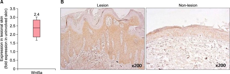
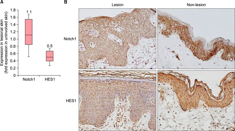
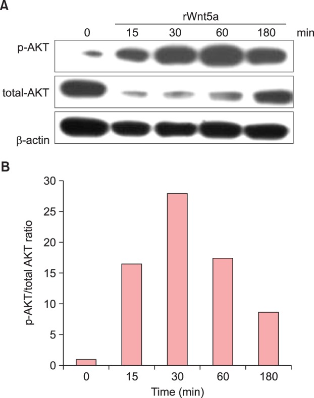
Results: In psoriatic lesions, expression of Wnt5a increased while that of Notch1 decreased when compared to that in non-lesional and normal skin. Treatment with rWnt5a increased the proliferation of keratinocytes and increased their secretion of interleukin (IL)-23, IL-12, and tumor necrosis factor (TNF)-α. Further, exposure of keratinocytes to IL-1α, TNF-α, transforming growth factor-α, and interferon-γ downregulated Notch1 as well as HES 1, which is downstream to Notch1, but increased the Wnt5a levels. The upregulated Wnt5a in keratinocytes downregulated both Notch1 and HES1.
Conclusion: Our data suggest that Wnt5a and Notch1 signaling exert counteracting influences on each other and are involved, in part, in the pathomechanism of psoriasis.
Keywords: Notch1; Psoriasis; Wnt5a.
Figures


Similar articles
-
Triptolide alleviates psoriasis through inhibiting the Wnt5a/β-Catenin signaling pathway.Front Pharmacol. 2025 Apr 29;16:1534118. doi: 10.3389/fphar.2025.1534118. eCollection 2025. Front Pharmacol. 2025. PMID: 40365308 Free PMC article.
-
Effect of γ-secretase inhibitor on Th17 cell differentiation and function of mouse psoriasis-like skin inflammation.J Transl Med. 2018 Mar 10;16(1):59. doi: 10.1186/s12967-018-1442-6. J Transl Med. 2018. PMID: 29523162 Free PMC article.
-
Biological therapy induces expression changes in Notch pathway in psoriasis.Arch Dermatol Res. 2015 Dec;307(10):863-73. doi: 10.1007/s00403-015-1594-7. Epub 2015 Aug 30. Arch Dermatol Res. 2015. PMID: 26319047
-
Beta-catenin non-canonical pathway: A potential target for inflammatory and hyperproliferative state via expression of transglutaminase 2 in psoriatic skin keratinocyte.Dermatol Ther. 2020 Nov;33(6):e14209. doi: 10.1111/dth.14209. Epub 2020 Sep 7. Dermatol Ther. 2020. PMID: 32816372 Review.
-
Intralesional T-lymphocyte activation as a mediator of psoriatic epidermal hyperplasia.J Invest Dermatol. 1995 Jul;105(1 Suppl):89S-94S. doi: 10.1111/1523-1747.ep12316121. J Invest Dermatol. 1995. PMID: 7616005 Review.
Cited by
-
The Wnt Blows: On the Functional Role of Wnt Signaling in Mycobacterium tuberculosis Infection and Beyond.Front Immunol. 2016 Dec 26;7:635. doi: 10.3389/fimmu.2016.00635. eCollection 2016. Front Immunol. 2016. PMID: 28082976 Free PMC article. Review.
-
The pathological role of Wnt5a in psoriasis and psoriatic arthritis.J Cell Mol Med. 2019 Sep;23(9):5876-5883. doi: 10.1111/jcmm.14531. Epub 2019 Jul 16. J Cell Mol Med. 2019. PMID: 31313518 Free PMC article. Review.
-
The value of WNT5A as prognostic and immunological biomarker in pan-cancer.Ann Transl Med. 2022 Apr;10(8):466. doi: 10.21037/atm-22-1317. Ann Transl Med. 2022. PMID: 35571400 Free PMC article.
-
Triptolide alleviates psoriasis through inhibiting the Wnt5a/β-Catenin signaling pathway.Front Pharmacol. 2025 Apr 29;16:1534118. doi: 10.3389/fphar.2025.1534118. eCollection 2025. Front Pharmacol. 2025. PMID: 40365308 Free PMC article.
-
Biomechanical Factors in Psoriatic Disease: Defective Repair Exertion as a Potential Cause. Hypothesis Presentation and Literature Review.ACR Open Rheumatol. 2019 Aug 8;1(7):452-461. doi: 10.1002/acr2.11056. eCollection 2019 Sep. ACR Open Rheumatol. 2019. PMID: 31777825 Free PMC article. Review.
References
-
- Nestle FO, Kaplan DH, Barker J. Psoriasis. N Engl J Med. 2009;361:496–509. - PubMed
-
- Reischl J, Schwenke S, Beekman JM, Mrowietz U, Stürzebecher S, Heubach JF. Increased expression of Wnt5a in psoriatic plaques. J Invest Dermatol. 2007;127:163–169. - PubMed
-
- Nicolas M, Wolfer A, Raj K, Kummer JA, Mill P, van Noort M, et al. Notch1 functions as a tumor suppressor in mouse skin. Nat Genet. 2003;33:416–421. - PubMed
LinkOut - more resources
Full Text Sources
Other Literature Sources
