

Non-invasive diagnostic techniques in the diagnosis of squamous cell carcinoma
- PMID: 26848316
- PMCID: PMC4733351
- DOI: 10.3315/jdcr.2015.1221
Non-invasive diagnostic techniques in the diagnosis of squamous cell carcinoma
Abstract
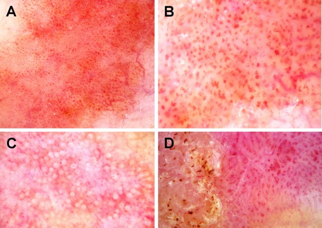
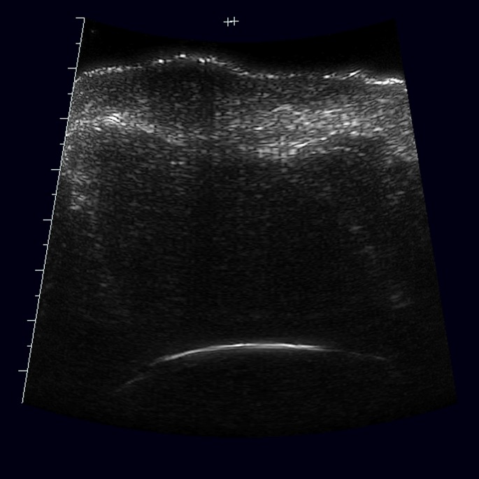
Squamous cell carcinoma is the second most common cutaneous malignancy after basal cell carcinoma. Although the gold standard of diagnosis for squamous cell carcinoma is biopsy followed by histopathology evaluation, optical non-invasive diagnostic tools have obtained increased attention. Dermoscopy has become one of the basic diagnostic methods in clinical practice. The most common dermoscopic features of squamous cell carcinoma include clustered vascular pattern, glomerular vessels and hyperkeratosis. Under reflectance confocal microscopy, squamous cell carcinoma shows an atypical honeycomb or disarranged pattern of the spinous-granular layer of the epidermis, round nucleated bright cells in the epidermis and round vessels in the dermis. High frequency ultrasound and optical coherence tomography may be helpful in predominantly in pre-surgical evaluation of tumor size. Emerging non-invasive or minimal invasive techniques with possible application in the diagnosis of squamous cell carcinoma of the skin, lip, oral mucosa, vulva or other tissues include high-definition optical coherence tomography, in vivo multiphoton tomography, direct oral microscopy, electrical impedance spectroscopy, fluorescence spectroscopy, Raman spectroscopy, elastic scattering spectroscopy, differential path-length spectroscopy, nuclear magnetic resonance spectroscopy, and angle-resolved low coherence interferometry.
Keywords: RCM; actinic keratosis; dermoscopy; diagnosis; oral mucous membrane; skin cancer; spectroscopy; squamous cell carcinoma; ultrasonography; vulva.
Figures

Similar articles
-
Role of In Vivo Reflectance Confocal Microscopy in the Analysis of Melanocytic Lesions.Acta Dermatovenerol Croat. 2018 Apr;26(1):64-67. Acta Dermatovenerol Croat. 2018. PMID: 29782304 Review.
-
Reflectance confocal microscopy criteria for squamous cell carcinomas and actinic keratoses.Arch Dermatol. 2009 Jul;145(7):766-72. doi: 10.1001/archdermatol.2009.134. Arch Dermatol. 2009. PMID: 19620557
-
Imaging actinic keratosis by high-definition optical coherence tomography. Histomorphologic correlation: a pilot study.Exp Dermatol. 2013 Feb;22(2):93-7. doi: 10.1111/exd.12074. Epub 2013 Jan 10. Exp Dermatol. 2013. PMID: 23301958
-
Non-invasive monitoring of subclinical and clinical actinic keratosis of face and scalp under topical treatment with ingenol mebutate gel 150 mcg/g by means of reflectance confocal microscopy and optical coherence tomography: New perspectives and comparison of diagnostic techniques.J Biophotonics. 2019 Jul;12(7):e201800391. doi: 10.1002/jbio.201800391. Epub 2019 Mar 20. J Biophotonics. 2019. PMID: 30653833
-
Minimally invasive modalities for keratinocyte carcinomas Part I: Diagnostics.J Am Acad Dermatol. 2025 Feb 21:S0190-9622(25)00319-6. doi: 10.1016/j.jaad.2025.02.036. Online ahead of print. J Am Acad Dermatol. 2025. PMID: 39993574 Review.
Cited by
-
The effect of wool hydrolysates on squamous cell carcinoma cells in vitro. Possible implications for cancer treatment.PLoS One. 2017 Aug 31;12(8):e0184034. doi: 10.1371/journal.pone.0184034. eCollection 2017. PLoS One. 2017. PMID: 28859143 Free PMC article.
-
Raman spectral post-processing for oral tissue discrimination - a step for an automatized diagnostic system.Biomed Opt Express. 2017 Oct 24;8(11):5218-5227. doi: 10.1364/BOE.8.005218. eCollection 2017 Nov 1. Biomed Opt Express. 2017. PMID: 29188115 Free PMC article.
-
Clinico-Pathologic Profile of a Cohort of Patients with Actinic Keratosis in a Tertiary Center in Romania.Cancers (Basel). 2025 Jun 10;17(12):1923. doi: 10.3390/cancers17121923. Cancers (Basel). 2025. PMID: 40563573 Free PMC article.
-
From Normal Skin to Squamous Cell Carcinoma: A Quest for Novel Biomarkers.Dis Markers. 2016;2016:4517492. doi: 10.1155/2016/4517492. Epub 2016 Aug 23. Dis Markers. 2016. PMID: 27642215 Free PMC article. Review.
-
Cutaneous Squamous Cell Carcinoma: Clinico-Dermoscopic and Histological Correlation: About 72 Cases.Dermatol Pract Concept. 2024 Jan 1;14(1):e2024042. doi: 10.5826/dpc.1401a42. Dermatol Pract Concept. 2024. PMID: 38364377 Free PMC article.
References
-
- Madan V, Lear JT, Szeimies RM. Non-melanoma skin cancer. Lancet. 2010;375:673–685. - PubMed
-
- Lomas A, Leonardi-Bee J, Bath-Hextall F. A systematic review of worldwide incidence of nonmelanoma skin cancer. Br J Dermatol. 2012;166:1069–1080. - PubMed
-
- Kauvar AN, Arpey CJ, Hruza G, Olbricht SM, Bennett R. Consensus for Nonmelanoma Skin Cancer Treatment, Part II: Squamous Cell Carcinoma, Including a Cost Analysis of Treatment Methods. Dermatol Surg. 2015;41:1214–1240. - PubMed
-
- Röwert-Huber J, Patel MJ, Forschner T, Ulrich C, Eberle J, Kerl H, Sterry W, Stockfleth E. Actinic keratosis is an early in situ squamous cell carcinoma: a proposal for reclassification. Br J Dermatol. 2007;156 Suppl 3:8–12. - PubMed
-
- Saphier J. Die Dermatoskopie.I. Mitteilung. Arch Dermatolo Syphiol. 1920;128:1–19.
Publication types
LinkOut - more resources
Full Text Sources
Other Literature Sources