

A rapidly growing giant cutaneous horn on the chest
- PMID: 26848321
- PMCID: PMC4733356
- DOI: 10.3315/jdcr.2015.1217
A rapidly growing giant cutaneous horn on the chest
Abstract
Background: A giant cutaneous horn (GCH) is a morphologic description of conical lesion with a dense, hyperkeratotic protrusion of more than 1 cm in height that resembles an animal horn but without its bony core. These can occur in association with benign, premalignant or malignant cutaneous diseases which can be determined by excision and histopathologic review of the base. A PubMed search (performed June 2015) revealed 54 cases of giant cutaneous horns in world literature. The most common site affected was the scalp followed by lip and leg. The commonest histological diagnosis found was squamous cell carcinoma followed by verruca vulgaris and trichilemmal horns.
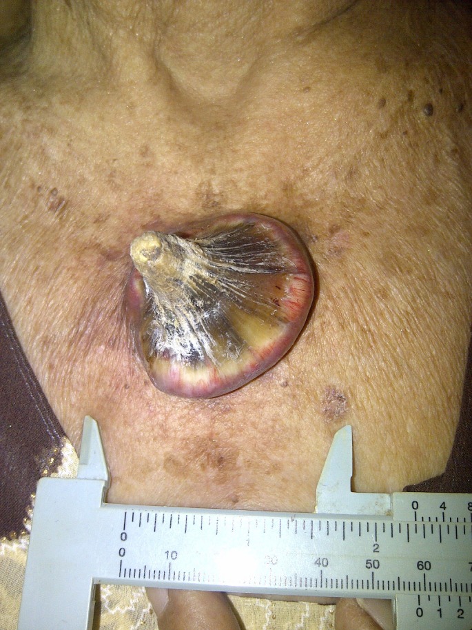
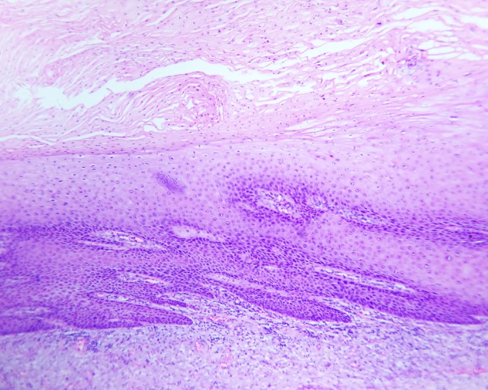
Main observation: We present an 85-year-old Filipino female with a one year history of a rapidly growing skin lesion on her upper chest. This was excised fully and histological review of the base demonstrated a keratoacanthoma.
Conclusions: This is the first known occurrence of a giant cutaneous horn on the chest. While giant cutaneous horns are more commonly associated with malignant lesions, differential diagnosis includes benign lesions such as keratoacanthomas. This differential can be considered in a rapidly growing lesion. Excision and histopathologic review of the base of a cutaneous horn are essential to guide potential further therapy.
Keywords: chest; cutaneous horn; keratoacanthoma; keratosis; skin cancer.
Figures
References
-
- Farris G. Histological considerations on a case of a voluminous cutaneous horn. Minerva Dermatol. 1953;28:159–165. - PubMed
-
- Michal M, Bisceglia M, Di Mattia A, Requena L, Fanburg-Smith JC, Mukensnabl P, Hes O, Cada F. Gigantic cutaneous horns of the scalp: lesions with a gross similarity to the horns of animals: a report of four cases. Am J Surg Pathol. 2002;26:789–794. - PubMed
Publication types
LinkOut - more resources
Full Text Sources
Other Literature Sources