

Cephalometric Angular Measurements of the Mandible Using Three-Dimensional Computed Tomography Scans in Koreans
- PMID: 26848443
- PMCID: PMC4738126
- DOI: 10.5999/aps.2016.43.1.32
Cephalometric Angular Measurements of the Mandible Using Three-Dimensional Computed Tomography Scans in Koreans
Abstract
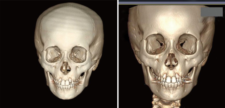
Background: We conducted this study to analyze the values of the key cephalometric angular measurements of the mandible using 3-dimensional (3D) computed tomography scans.
Methods: In the 106 enrolled patients, a 3D cephalometric analysis was performed to measure the angular variables of the mandible. These values were compared between the two sides and between the two sexes.
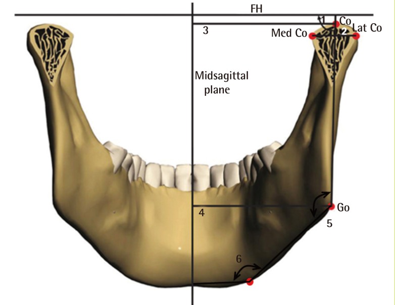
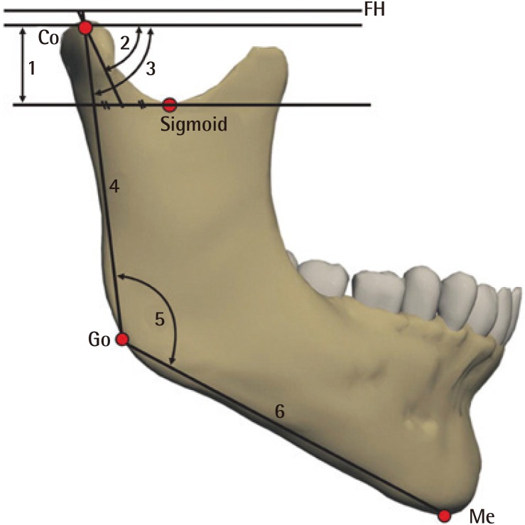
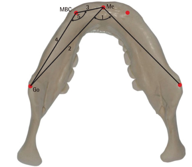
Results: The frontal measurements revealed that the mandibular body curve angle was larger on the left (Lt) side (right [Rt], 141.24±7.54; Lt, 142.68±6.94; P=0.002) and the gonial angle was larger on the right side (Rt, 134.37±8.44; Lt, 131.54±7.14; P<0.001). The sagittal measurements showed that the gonial angle was larger on the right side (Rt, 134.37±8.44; Lt, 131.54±7.14; P>0.05). Further, the transverse measurements revealed that the mandibular body curve angle was larger on the right side (Rt, 140.28±7.05; Lt, 137.56±6.23; P<0.001).
Conclusions: These results provide an average of the mandibular angular measurements for the Korean population, establishing a standard for determining surgical patient groups and outcome evaluations in the field of mandible contour surgery.
Keywords: Angular; Cephalometry; Mandible; Three-dimensional.
Conflict of interest statement
No potential conflict of interest relevant to this article was reported.
Figures
References
-
- Pacini SJ. Roentgen ray anthropometry of the skull. J Radiol. 1922;3:230–238.
-
- Broadbent BH. A new x-ray technique and its application to orthodontia. Angle Orthod. 1931;1:45–66.
LinkOut - more resources
Full Text Sources
Other Literature Sources