

Novel Acellular Scaffold Made from Decellularized Schwann Cell Sheets for Peripheral Nerve Regeneration
- PMID: 26848489
- PMCID: PMC4734393
- DOI: 10.1007/s40883-015-0003-2
Novel Acellular Scaffold Made from Decellularized Schwann Cell Sheets for Peripheral Nerve Regeneration
Abstract
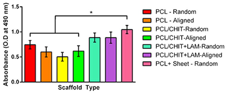
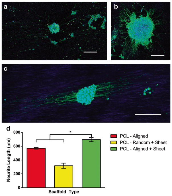
Extracellular matrix surrounding Schwann cells and neurons provides critical determinants of cellular phenotype during development as well as essential cues in stimulating and guiding regrowth. Using cell sheet technology, we developed a novel scaffold enriched with native extracellular matrix from Schwann cells. Schwann cells were grown into sheets and layered onto polycaprolactone fibers for support. Upon decellularization of these constructs, extracellular matrix remained with few traces of nucleic acids. This method of deposition of extracellular matrix provided more protein than traditional seeding method after decellularization. Additionally, the isolated matrix supported proliferation of Schwann cells better than covalently bound laminin. The proliferation and differentiation of Schwann cells grown on decellularized sheets were complemented by upregulation of Erbb2 and myelin protein zero. Laminin expression of β1 and γ1 chains was also elevated. PC12 cells grown on decellularized sheets produced longer neurite extensions than aligned polycaprolactone fibers alone, proving potential of these scaffolds to be used in future peripheral nerve regenerative studies.
Lay summary: Peripheral nerve injuries present a serious clinical need with approximately 50 % of surgical cases achieving only some restoration of function. In order to better guide regenerating nerves, supporting cells of the nerve tissue were grown into sheets and subsequently decellularized, leaving a myriad of surrounding protein as a scaffold. Constructs have been shown to support cell growth and neurite extension in vitro. Future projects will combine various cell types present in the nerve tissue as well as stem cells to fully support and reconstruct architecture of the peripheral nerves.
Keywords: Cell sheet; Decellularization; Extracellular matrix; Nerve tissue engineering; Schwann cell.
Figures


Similar articles
-
Biocompatibility evaluation of electrospun aligned poly (propylene carbonate) nanofibrous scaffolds with peripheral nerve tissues and cells in vitro.Chin Med J (Engl). 2011 Aug;124(15):2361-6. Chin Med J (Engl). 2011. PMID: 21933569
-
Novel Sodium Deoxycholate-Based Chemical Decellularization Method for Peripheral Nerve.Tissue Eng Part C Methods. 2020 Jan;26(1):23-36. doi: 10.1089/ten.TEC.2019.0135. Epub 2019 Dec 19. Tissue Eng Part C Methods. 2020. PMID: 31724493
-
Cultures of Schwann-like cells differentiated from adipose-derived stem cells on PDMS/MWNT sheets as a scaffold for peripheral nerve regeneration.J Biomed Mater Res A. 2015 Nov;103(11):3642-8. doi: 10.1002/jbm.a.35488. Epub 2015 Jun 25. J Biomed Mater Res A. 2015. PMID: 25903927
-
Therapeutic strategies for peripheral nerve injury: decellularized nerve conduits and Schwann cell transplantation.Neural Regen Res. 2019 Aug;14(8):1343-1351. doi: 10.4103/1673-5374.253511. Neural Regen Res. 2019. PMID: 30964052 Free PMC article. Review.
-
Expression and functional roles of neural cell surface molecules and extracellular matrix components during development and regeneration of peripheral nerves.J Neurocytol. 1994 Jan;23(1):1-28. doi: 10.1007/BF01189813. J Neurocytol. 1994. PMID: 8176415 Review.
Cited by
-
Fabrication and Applications of Micro/Nanostructured Devices for Tissue Engineering.Nanomicro Lett. 2017;9(1):1. doi: 10.1007/s40820-016-0103-7. Epub 2016 Aug 31. Nanomicro Lett. 2017. PMID: 30460298 Free PMC article. Review.
-
Comparing Processed Nerve Allografts and Assessing Their Capacity to Retain and Release Nerve Growth Factor.Ann Plast Surg. 2018 Aug;81(2):198-202. doi: 10.1097/SAP.0000000000001464. Ann Plast Surg. 2018. PMID: 29781850 Free PMC article.
-
Transplantation of Neural Progenitor Cells Derived from Stem Cells from Apical Papilla Through Small-Molecule Induction in a Rat Model of Sciatic Nerve Injury.Tissue Eng Regen Med. 2024 Aug;21(6):867-879. doi: 10.1007/s13770-024-00648-y. Epub 2024 Jun 21. Tissue Eng Regen Med. 2024. PMID: 38904732 Free PMC article.
-
Design of Novel Mechanically Resistant and Biodegradable Multichannel Platforms for the Treatment of Peripheral Nerve Injuries.Biomacromolecules. 2023 Apr 10;24(4):1731-1743. doi: 10.1021/acs.biomac.2c01498. Epub 2023 Mar 15. Biomacromolecules. 2023. PMID: 36922716 Free PMC article.
-
Fabrication and Evaluation of Porous dECM/PCL Scaffolds for Bone Tissue Engineering.J Funct Biomater. 2023 Jun 29;14(7):343. doi: 10.3390/jfb14070343. J Funct Biomater. 2023. PMID: 37504838 Free PMC article.
References
Grants and funding
LinkOut - more resources
Full Text Sources
Other Literature Sources
Research Materials
Miscellaneous