

Review
doi: 10.1097/RMR.0000000000000073.
Presurgical Mapping of the Language Network Using Resting-state Functional Connectivity
Affiliations
- PMID: 26848557
- PMCID: PMC4833007
- DOI: 10.1097/RMR.0000000000000073
Item in Clipboard
Review
Presurgical Mapping of the Language Network Using Resting-state Functional Connectivity
Top Magn Reson Imaging.
2016 Feb.
Abstract
Resting-state functional magnetic resonance imaging (resting-state fMRI) is a tool for investigating the functional networks that arise during the resting state of the brain. Recent advances of the resting-state fMRI analysis suggest its feasibility for evaluating language function. The most common clinical application is for presurgical mapping of cortex for a brain tumor or for resective epilespy surgery. In this article, we review the techniques and presurgical applications of resting-state fMRI analysis for language evaluation, and discuss the use in the clinical setting, focusing on planning for neurosurgery.
Figures
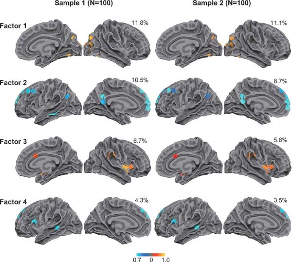
Factor analysis derived from the resting-fMRI intrinsic connectivity shows four significant clusters, each of which represents the visual system (Factor 1), default mode network (Factor 2), attention system (Factor 3) and language network (Factor 4). Used with permission (17)
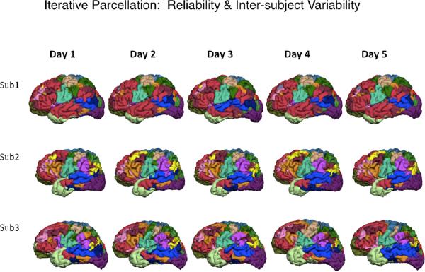
The parcellation atlas of individuals show the intra-subject reliability and inter-subject variability of the functional networks. (Courtesy of Hesheng Liu, Ph. D)
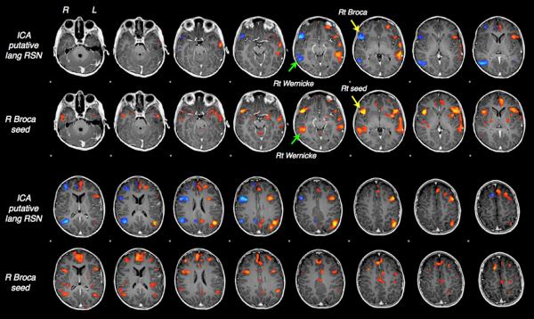
ICA-based mapping of the language network, showing the Wernicke and Broca areas consistently with the results of seed-based approach. (Courtesy of Brad Buchbinder, M.D)
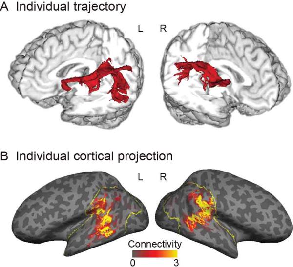
A. Tractography represents the arcuate fasciculus. B. Cortical projection of tractography calculated by the surface-based analysis. Used with permission (29).
Similar articles
-
Language Mapping Using fMRI and Direct Cortical Stimulation for Brain Tumor Surgery: The Good, the Bad, and the Questionable.Top Magn Reson Imaging. 2016 Feb;25(1):1-10. doi: 10.1097/RMR.0000000000000074. Top Magn Reson Imaging. 2016. PMID: 26848555 Free PMC article. Review.
-
Resting-state functional MRI in pediatric epilepsy surgery.Pediatr Neurosurg. 2013;49(5):261-73. doi: 10.1159/000363605. Epub 2014 Sep 24. Pediatr Neurosurg. 2013. PMID: 25277135 Review.
-
Real-time presurgical resting-state fMRI in patients with brain tumors: Quality control and comparison with task-fMRI and intraoperative mapping.Hum Brain Mapp. 2020 Feb 15;41(3):797-814. doi: 10.1002/hbm.24840. Epub 2019 Nov 6. Hum Brain Mapp. 2020. PMID: 31692177 Free PMC article.
-
Presurgical brain mapping of the language network in pediatric patients with epilepsy using resting-state fMRI.J Neurosurg Pediatr. 2021 Jan 8;27(3):259-268. doi: 10.3171/2020.8.PEDS20517. Print 2021 Mar 1. J Neurosurg Pediatr. 2021. PMID: 33418528
-
Clinical Resting-state fMRI in the Preoperative Setting: Are We Ready for Prime Time?Top Magn Reson Imaging. 2016 Feb;25(1):11-8. doi: 10.1097/RMR.0000000000000075. Top Magn Reson Imaging. 2016. PMID: 26848556 Free PMC article. Review.
Cited by
-
Role of Functional Imaging Techniques to Assess Motor and Language Cortical Plasticity in Glioma Patients: A Systematic Review.Neural Plast. 2019 Nov 11;2019:4056436. doi: 10.1155/2019/4056436. eCollection 2019. Neural Plast. 2019. PMID: 31814822 Free PMC article.
-
Language processing in Internet use disorder: Task-based fMRI study.PLoS One. 2022 Jun 24;17(6):e0269979. doi: 10.1371/journal.pone.0269979. eCollection 2022. PLoS One. 2022. PMID: 35749379 Free PMC article.
-
Functional MRI for Surgery of Gliomas.Curr Treat Options Neurol. 2017 Aug 23;19(10):34. doi: 10.1007/s11940-017-0469-y. Curr Treat Options Neurol. 2017. PMID: 28831723 Review.
-
Mapping language function with task-based vs. resting-state functional MRI.PLoS One. 2020 Jul 31;15(7):e0236423. doi: 10.1371/journal.pone.0236423. eCollection 2020. PLoS One. 2020. PMID: 32735611 Free PMC article.
-
Determination of Differences in Seed-Based Resting State Functional Magnetic Resonance Imaging Language Networks in Pediatric Patients with Left- and Right-Lateralized Language: A Pilot Study.J Epilepsy Res. 2019 Dec 31;9(2):93-102. doi: 10.14581/jer.19011. eCollection 2019 Dec. J Epilepsy Res. 2019. PMID: 32509544 Free PMC article.
References
-
- Dion JE, Gates PC, Fox AJ, et al. Clinical events following neuroangiography: a prospective study. Stroke. 1987;18:997–1004. - PubMed
-
- Snyder PJ, Harris LJ. The intracarotid amobarbital procedure: an historical perspective. Brain Cogn. 1997;33:18–32. - PubMed
-
- De Paola L, Mader MJ, Germiniani FM, et al. Bizarre behavior during intracarotid sodium amytal testing (WADA test): are they predictable? Arq Neuropsiquiatr. 2004;62:444–8. - PubMed
-
- Dym RJ, Burns J, Freeman K, et al. Is functional MR imaging assessment of hemispheric language dominance as good as the Wada test?: a meta-analysis. Radiology. 2011;261:446–55. - PubMed
Publication types
MeSH terms
Grants and funding
LinkOut - more resources
Full Text Sources
Other Literature Sources
Medical
