Review
. 2016 Feb;25(1):25-30.
doi: 10.1097/RMR.0000000000000077.

Cortical Plasticity in the Setting of Brain Tumors

Affiliations
Review

Cortical Plasticity in the Setting of Brain Tumors

Ryan A Fisicaro et al. Top Magn Reson Imaging. 2016 Feb.

Abstract

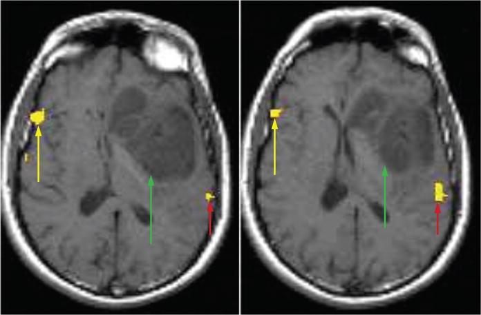
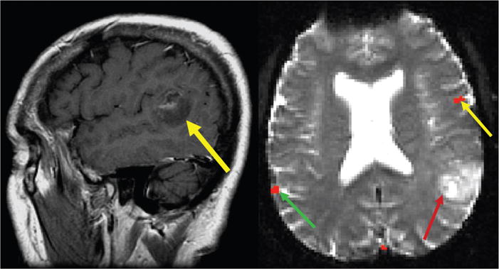
Cortical reorganization of function due to the growth of an adjacent brain tumor has clearly been demonstrated in a number of surgically proven cases. Such cases demonstrate the unmistakable implications for the neurosurgical treatment of brain tumors, as the cortical function may not reside where one may initially suspect based solely on the anatomical magnetic resonance imaging (MRI). Consequently, preoperative localization of eloquent areas adjacent to a brain tumor is necessary, as this may demonstrate unexpected organization, which may affect the neurosurgical approach to the lesion. However, in interpreting functional MRI studies, the interpreting physician must be cognizant of artifacts, which may limit the accuracy of functional MRI in the setting of brain tumors.


Figures

FIGURE 1
FIGURE 1
fMRI scan with language paradigms coregistered to axial T1-weighted images in a 34-year-old man with a left inferior frontal glioma. The 2 axial slices localize Broca area to the right hemisphere (yellow arrows), Wernicke area to the left hemisphere (red arrows), and show their relationship to the tumor location (green arrows). The localization of Broca area to the right hemisphere suggests cortical reorganization. Axial slices taken from a figure in J Comput Assist Tomogr 2002; 26:941–943.
FIGURE 2
FIGURE 2
A, Postcontrast sagittal image from a 62-year-old right-handed man with a mostly nonenhancing temporoparietal neoplasm involving the expected location of Wernicke area (yellow arrow). B, Axial fMRI results show Broca area localizing to the left hemisphere (yellow arrow) and Wernicke area localizing to the right hemisphere (green arrow) opposite of the tumor (red arrow). Intraoperative mapping and surgical resection confirmed fMRI results. Adapted from AJNR Am J Neuroradiol 2004; 25:130–133.
FIGURE 3
FIGURE 3
A patient with a GBM in the left motor cortex performing a bilateral finger tapping paradigm. The top graphs (A) are the response curves of finger tapping on the right hand (which are mapped to the tumor-affected hemisphere) and the bottom graphs (B) are the response curves of finger tapping on the left hand (which are mapped to the normal, contralateral hemisphere). In A, we see that that the peak in the hemodynamic response function curve occurs 0.5 seconds sooner in the SMA than the motor cortex, while in B, the SMA peak 1.5 seconds sooner than in the motor cortex. The shift in the SMA peak in the tumor-affected hemisphere suggests that the SMA may be taking on some motor functions from the motor cortex. Used and adapted with permission from Med Sci Monit 2009; 15: MT55–MT62.

Similar articles

Cited by

References

    1. Bailey CH, Kandel ER, Harris KM. Structural components of synaptic plasticity and memory consolidation. Cold Spring Harb Perspect Biol. 2015;7:a021758. - PMC - PubMed
    1. Kandel ER, Dudai Y, Mayford MR. The molecular and systems biology of memory. Cell. 2014;157:163–186. - PubMed
    1. Temple E, Deutsch GK, Poldrack RA, et al. Neural deficits in children with dyslexia ameliorated by behavioral remediation: evidence from functional MRI. Proc Natl Acad Sci U S A. 2003;100:2860–2865. - PMC - PubMed
    1. Lind J, Persson J, Ingvar M, et al. Reduced functional brain activity response in cognitively intact apolipoprotein E epsilon4 carriers. Brain. 2006;129(pt 5):1240–1248. - PubMed
    1. Trivedi MA, Schmitz TW, Ries ML, et al. Reduced hippocampal activation during episodic encoding in middle-aged individuals at genetic risk of Alzheimer’s disease: a cross-sectional study. BMC Med. 2006;4:1. - PMC - PubMed

MeSH terms