. 2016 May;14(5):1082-94.
doi: 10.1111/jth.13281. Epub 2016 Mar 22.

Non-genomic activities of retinoic acid receptor alpha control actin cytoskeletal events in human platelets

Affiliations

Non-genomic activities of retinoic acid receptor alpha control actin cytoskeletal events in human platelets

M T Rondina et al. J Thromb Haemost. 2016 May.

Abstract

Essentials Platelets employ proteins/signaling pathways traditionally thought reserved for nuclear niche. We determined retinoic-acid-receptor alpha (RARα) expression and function in human platelets. RARα/actin-related protein-2/3 complex (Arp2/3) interact via non-genomic signaling in platelets. RARα regulates Arp2/3-mediated actin cytoskeletal dynamics and platelet spreading.

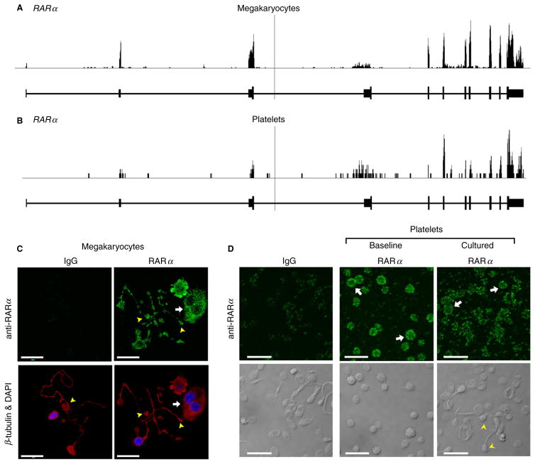
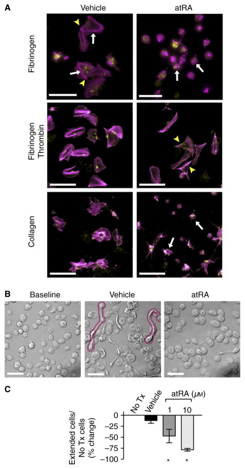
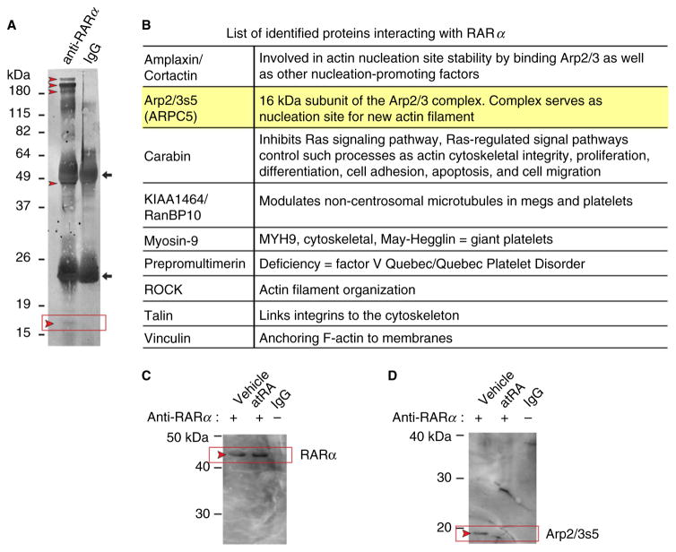
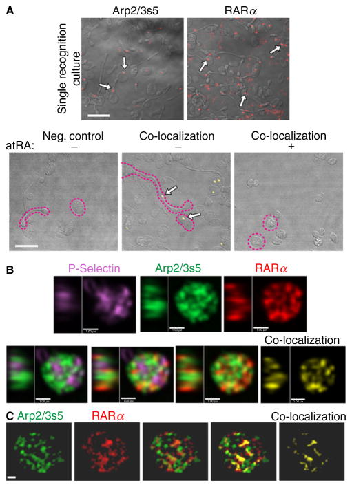
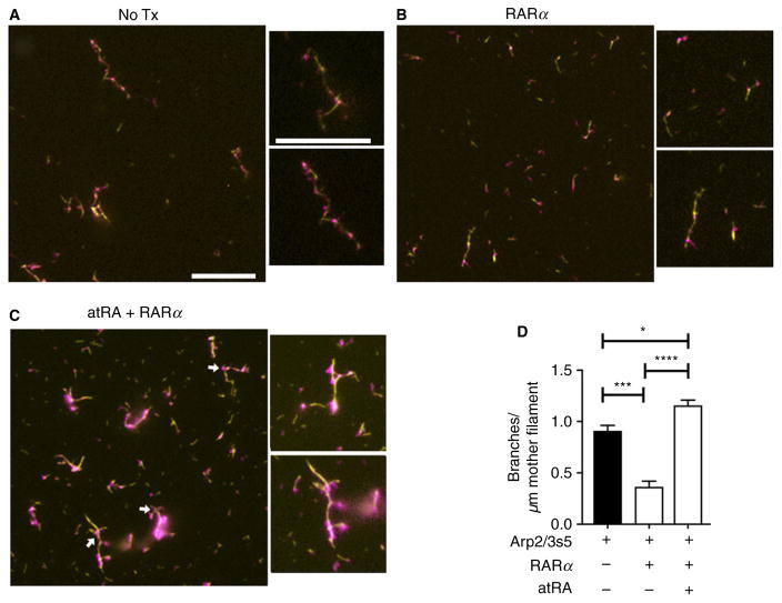
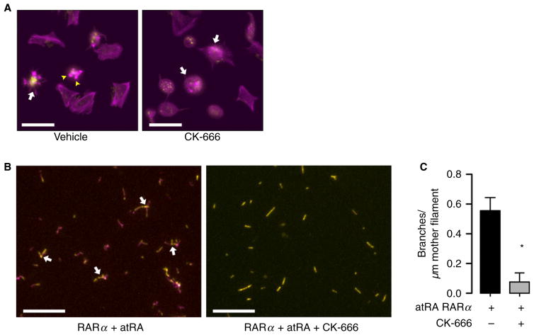
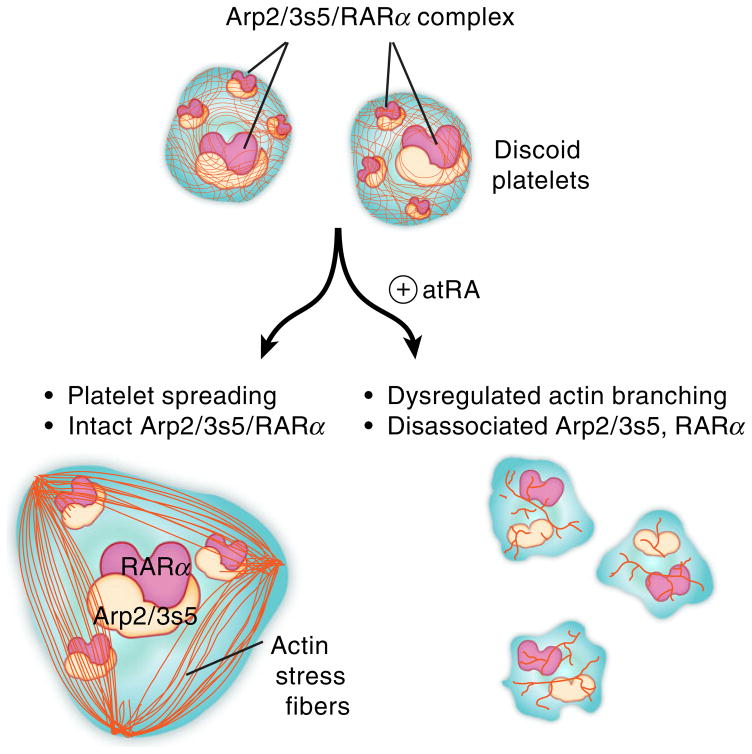
Summary: Background Platelets utilize proteins and pathways classically reserved for the nuclear niche. Methods We determined whether human platelets express retinoic-acid-receptor family members, traditionally thought of as nuclear transcription factors, and deciphered the function of RARα. Results We found that RARα is robustly expressed in human platelets and megakaryocytes and interacts directly with actin-related protein-2/3 complex (Arp2/3) subunit 5 (Arp2/3s5). Arp2/3s5 co-localized with RARα in situ and regulated platelet cytoskeletal processes. The RARα ligand all-trans retinoic acid (atRA) disrupted RARα-Arp2/3 interactions. When isolated human platelets were treated with atRA, rapid cytoskeletal events (e.g. platelet spreading) were inhibited. In addition, when platelets were cultured for 18 h in the presence of atRA, actin-dependent morphological changes (e.g. extended cell body formation) were similarly inhibited. Using in vitro actin branching assays, RARα and Arp2/3-regulated complex actin branch formation was demonstrated. Consistent with inhibition of cytoskeletal processes in platelets, atRA, when added to this branching assay, resulted in dysregulated actin branching. Conclusion Our findings identify a previously unknown mechanism by which RARα regulates Arp2/3-mediated actin cytoskeletal dynamics through a non-genomic signaling pathway. These findings have broad implications in both nucleated and anucleate cells, where actin cytoskeletal events regulate cell morphology, movement and division.

Keywords: actin; actin-related protein 2-3 complex; blood platelets; protein interaction domains and motifs; retinoic acid receptors.


Conflict of interest statement

Disclosure of Conflict of Interests

The authors state that they have no conflict of interest.

Figures

Fig. 1
Fig. 1
Megakaryocytes and human platelets express mRNA and protein for RARα. RNA-seq snapshots taken from the Integrated Genome Browser of the RARα transcript in CD34+-derived megakaryocytes (A) and human platelets (B). The height on the y-axis represents the relative accumulated number of reads spanning a particular sequence. The average of the read depths across all genomic coordinates within a transcript correlates to abundance of RNA expression. RAR gene regions are represented below the plots by thick (exon) and thin (intron) lines. (C) CD34+-derived megakaryocytes were placed on immobilized fibrinogen to induce proplatelet formation, fixed, and then incubated with an antibody against RARα (green, top right panel) or an IgG control, followed by co-staining for β-tubulin (red) and nuclei (4′,6-diamidino-2-phenylindole, DAPI, blue), as shown in the bottom panels. The white arrows point to cell bodies and yellow arrowheads highlight the proplatelet extensions. Scale bars = 20 μm. (D) RARα expression (green) at baseline and in cultured (18 h) human platelets. The bottom panels show the corresponding transmission images. The white arrows highlight RARα platelets. The yellow arrowheads point to barbell-shaped platelets that form when platelets are cultured in suspension [4]. Scale bars = 10 μm.
Fig. 2
Fig. 2
Retinoic acid regulates platelet progeny formation. (A) Platelets were placed on immobilized fibrinogen (top), fibrinogen plus thrombin (middle) or collagen (bottom) for 2 h with vehicle (dimethylsulfoxide, DMSO) or in the presence of atRA (10 μM) and stained for polymerized actin (magenta) or wheat germ agglutinin (WGA) to identify sialic acids (yellow). In the presence of vehicle only, isolated human platelets demonstrate characteristic fully spread platelets and star-shaped, partially spread patterns with hallmark actin stress fibers (arrows) and actin nodules (arrowheads). Treatment with atRA inhibited full platelet spreading, although partially spread, star-shaped platelets remain visible (white arrows, scale bars = 10 μm). This figure is representative of n = 3 independent experiments. (B) Transmission microscopy of platelets following snap-fixation at baseline or following incubation (6 h) with vehicle (DMSO) or atRA (10 μM). The formation of extended platelets with ≥ 2 cell bodies (outlined in magenta dotted line) was reduced compared with baseline or vehicle-treated platelets. Scale bars = 10 μm, representative of n = 5 independent experiments. (C) Extended platelets with ≥ 2 cell bodies were quantified without treatment (no Tx) and during treatment in culture with either vehicle (DMSO) or atRA in a dose-dependent manner (1 and 10 μM). Data represent the mean ± SEM (n = 3, *P < 0.05).
Fig. 3
Fig. 3
Arp2/3s5, a subunit of the actin-nucleating complex that regulates cytoskeletal formation, is robustly expressed in human platelets and CD34+-derived megakaryocytes (MEGS) and interacts with RARα. (A) A poly(vinylidene difluoride) (PVDF) membrane stained for total protein (colloidal gold). The corresponding sodium dodecyl sulfate (SDS) gel stained with coomassie was used for mass spectroscopy (MS)-based protein identification. The left lane shows proteins isolated after co-immunoprecipitation (co-IP) using an anti-RARα antibody. The right lane shows proteins isolated after co-IP with non-immunogenic, control IgG. The IgG heavy and light chains are indicated with black arrows. Red arrowheads mark selected protein bands subsequently used in MS analysis. The expected position of ARP2/3s5 is indicated by a black box, between 15 and 19 kDa, where a faint but visible band is present. (B) List of identified proteins interacting with RARα. Protein names are shown in the left column, known protein functions are listed in the right column. Highlighted is our primary target (Arp2/3s5). (C, D) atRA blocks RARα Arp2/3s5 interactions. Platelets were cultured overnight in the absence (vehicle, left lane) or presence of atRA (10 μM, middle lane). Platelet lysates were then analyzed by co-IP with an anti-RARα antibody followed by Western blotting. The corresponding IgG control is shown in the right lane. (C) An anti-RARα antibody was used for the detection of immuno-precipitated RARα. (D) Membranes were stripped and reprobed for the presence of Arp2/3s5. The red arrowheads indicate the expected positions of RARα and Arp2/3s5 on the membrane. (C) and (D) are representative of n = 3 independent experiments.
Fig. 4
Fig. 4
RARα and Arp2/3s5 co-localize in situ. (A) Platelets were cultured overnight, fixed and spun down. Single recognition studies were performed using the Duolink® in situ system. RARα and Arp2/3s5 protein were detected using specific primary antibodies. Single recognition signals for either RARα or Arp2/3s5 (red, top panels) are highlighted in each panel with white arrows, demonstrating that both RARα and Arp2/3s5 are present within human platelets. Co-localization studies were performed using the Duolink® in situ system. RARα-Arp2/3s5 complexes were detected by using specific primary antibodies and matching (−) and (+) PLA probes. Co-localization signals for RARα Arp2/3s5 complexes (yellow, bottom panels) are identified by white arrows. The negative control (left panel) demonstrates specificity of the signal. Examples of discoid and extended cells are outlined (magenta dotted line). Scale bars = 10 μm. Images in (A) are representative of n = 3 independent experiments. (B and C) Human platelets were isolated, prepared and immunostained using primary antibodies: mouse anti-RARα (red), rabbit anti-Arp2/3s5 (green) and goat anti-P selectin (magenta). Stained cells were imaged via spinning disc confocal laser fluorescence microscopy (B) or structured illumination microscopy (SIM) (C). The top row panels in (B) and the two left side panels in (C) show single channel images for each protein indicated. Merged images used for co-localization studies are shown in the bottom row (B) and middle panels in (C). Co-localization pixels only for RARα and Arp2/3s5 are shown in the far right panels (B, C) in yellow. In (B) the left side in each panel shows the central YZ profile of the Z-section shown at right, in (C) images are snapshots of the 3D render shown in Movie S1 (B, bars = 1 μm, representative of three independent experiments; C, bars = 0.5 μm, representative of three independent experiments).
Fig. 5
Fig. 5
Retinoic acid receptor-α and atRA are required for complex actin branching in the presence of Arp2/3. An actin branching assay was performed as described in ‘Materials and methods’. Simple actin mother filaments are shown in yellow while complex actin branching is shown in magenta. (A) In the presence of the Arp2/3 complex and WASP-VCA alone (control), actin polymerization and actin branching were detectable. (B, D) When recombinant human RARα (100 nM final) was added, actin branching formation and the number of actin side-branches per μM of actin mother filament were significantly reduced. (C, D) In comparison, atRA significantly rescued actin branch formation, even in the presence of RARα (magenta, white arrows). Scale bars = 10 μM; these figures are representative of n = 3 independent experiments.
Fig. 6
Fig. 6
Actin cytoskeletal dynamics in human platelets is dependent on Arp2/3s5. (A) Platelets were placed on immobilized fibrinogen for 2 h with vehicle (dimethylsulfoxide, DMSO) or in the presence of CK-666, a specific Arp2/3s5 inhibitor, and stained for polymerized actin (magenta) or wheat germ agglutinin (WGA) to identify sialic acids (yellow). In the presence of vehicle only, isolated human platelets demonstrate characteristic fully spread platelets and star-shaped (arrows), partially spread patterns with hallmark actin stress fibers and focal adhesion points (arrowheads). Treatment with CK-666, in contrast, inhibited full platelet spreading, although partially spread, star-shaped platelets remain visible (white arrows, scale bars = 10 μm). This figure is representative of n = 3 independent experiments. (B, C) When an actin branching assay was supplemented in control conditions [18,19] with bovine Arp2/3 complex in the presence of recombinant human RARα and atRA, actin filaments were visible and complex actin branching occurred. In the presence of the Arp2/3 inhibitor CK-666, actin filaments were visible but complex actin branching was inhibited, consistent with the dependency of the Arp2/3 complex on actin branch formation. Boxes indicate the origin of inserts. Scale bars = 10 μm. This figure is representative of n = 3 independent experiments.
Fig. 7
Fig. 7
Proposed mechanisms by which retinoic acid regulates complex actin cytoskeletal dynamics in human platelets. Basally, in unstimulated human platelets, RARα is bound to Arp2/3s5 and these direct interactions mediate platelet shape change and spreading, processes dependent on regulated actin cytoskeletal events. These interactions are disrupted by the binding of retinoic acid to its receptor, leading to dysregulated actin cytoskeletal rearrangement and blocking of platelet shape change and spreading.

Similar articles

Cited by

References

    1. Hartwig JH. Mechanisms of actin rearrangements mediating platelet activation. J Cell Biol. 1992;118:1421–42. - PMC - PubMed
    1. Hartwig JH. The platelet: form and function. Semin Hematol. 2006;43:S94–100. - PubMed
    1. White JG. An overview of platelet structural physiology. Scanning Microsc. 1987;1:1677–700. - PubMed
    1. Schwertz H, Koster S, Kahr WH, Michetti N, Kraemer BF, Weitz DA, Blaylock RC, Kraiss LW, Greinacher A, Zimmerman GA, Weyrich AS. Anucleate platelets generate progeny. Blood. 2010;115:3801–9. - PMC - PubMed
    1. Thon JN, Montalvo A, Patel-Hett S, Devine MT, Richardson JL, Ehrlicher A, Larson MK, Hoffmeister K, Hartwig JH, Italiano JE., Jr Cytoskeletal mechanics of proplatelet maturation and platelet release. J Cell Biol. 2010;191:861–74. - PMC - PubMed

Publication types