. 2016 Mar 1;7(9):9680-91.
doi: 10.18632/oncotarget.7109.

Guanylate binding protein-1 mediates EGFRvIII and promotes glioblastoma growth in vivo but not in vitro

Affiliations

Guanylate binding protein-1 mediates EGFRvIII and promotes glioblastoma growth in vivo but not in vitro

Qing Lan et al. Oncotarget. .

Abstract

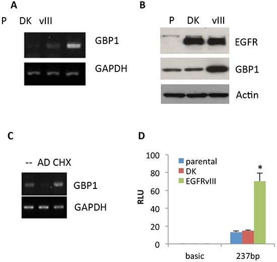
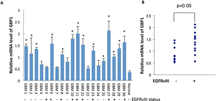
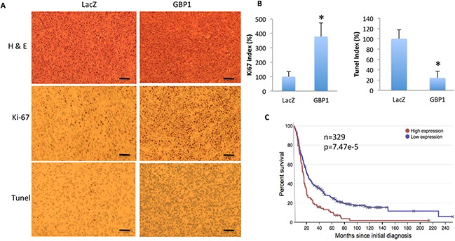
Glioblastoma multiforme (GBM) is the most common and deadly primary brain tumor in adults. Epidermal growth factor receptor (EGFR) is frequently amplified and mutated in GBM. We previously reported that Guanylate binding protein-1 (GBP1) is a novel transcriptional target gene of EGFR and plays a role in GBM invasion. Here we demonstrate that GBP1 can also be induced by EGFRvIII at the transcriptional level through the p38 MAPK/Yin Yang 1 (YY1) signaling pathway. Silencing of GBP1 by RNA interference significantly inhibits EGFRvIII-mediated GBM cell proliferation in vitro and in a mouse model. Overexpression of GBP1 has no obvious effect on glioblastoma cell proliferation in vitro. In contrast, in an orthotopic glioma mouse model GBP1 overexpression significantly promotes glioma growth and reduces survival rate of glioma-bearing mice by increasing cell proliferation and decreasing cell apoptosis in tumor. Clinically, GBP1 expression is elevated in human GBM tumors and positively correlates with EGFRvIII status in GBM specimens, and its expression is inversely correlated with the survival rate of GBM patients. Taken together, these results reveal that GBP1 may serve as a potential therapeutic target for GBMs with EGFRvIII mutation.

Keywords: EGFRvIII; GBP1; glioblastoma; survival; tumor growth.


Conflict of interest statement

CONFLICTS OF INTEREST

The authors declare no conflict of interest.

Figures

Figure 1
Figure 1. EGFRvIII promotes GBP1 expression
The expression of GBP1 was analyzed by semiquantitative RT-PCR A. and Western blot B. in U87-parental (p), U87-DK and U87-EGFRvIII cells which were serum starved for 48 hr. C. After 24 h of serum starvation, U87-P, U87-DK and U87-EGFRvIII cells were treated with AD or CHX for an additional 24 h. GBP1 and GAPDH mRNA was measured by RT-PCR. D. U87-P, U87-DK and U87-EGFRvIII cells were transfected with pGL3-237 and pRL-TK for 24 h and then serum starved for 24 h. Firefly and Renilla luciferase activities were measured, and promoter activity is presented as the fold induction of RLU (values of firefly luciferase unit/values of Renilla) as compared with control. The results are expressed as the mean of three independent experiments ± SD. *, P < 0.01.
Figure 2
Figure 2. GBP1 is upregulated and correlated with EGFRvIII status in GBM specimens
A. GBP1 expression in GBM patients was measured by RT-qPCR analysis. Normal denotes normal brain tissue. *, p < 0.05. B. Correlation analysis between GBP1 and EGFRvIII status in human GBM samples was analyzed for significance using the Kruskal Wallis test; P < 0.05 was considered statistically significant.
Figure 3
Figure 3. EGFRvIII–stimulated GBP1 expression is p38 MAPK dependent
A. After 24 h of serum starvation, U87-EGFRvIII cells were treated with DMSO (−), 10 μM of the EGFR tyrosine kinase inhibitor AG1478 (AG), or 20μM of the p38 inhibitor SB203580 (SB) for an additional 24 h before Western blot analysis. B. U87-EGFRvIII cells were transfected with the indicated concentration of p38 siRNA (si-p38) or control siRNA (si-Luc) for 24 h and then serum starved for 24 h followed by Western blot analysis. The p38 siRNAs were described previously [10]. C. U87-EGFRvIII cells were transfected with pGL3-237 and pRL-TK for 24 h and then serum starved for 24 h. The starved cells were pretreated with DMSO or 20 μM SB203580 for an additional 24 h before reporter assay. This result is expressed as the mean of three independent experiments ± SD. *, P < 0.01. D. Levels of GBP1, EGFRvIII and phospho-p38 were assessed by Western blot analysis in U87 cells expressing low, medium and high levels of EGFRvIII, which were generated as described elsewhere [25].
Figure 4
Figure 4. YY1 is involved in regulation of EGFRvIII-mediated GBP1 expression
A. U87-EGFRvIII cells were transfected with the GBP1 wild-type promoter pGL3-237 or the YY1 deactivated promoter pGL3-237-YY1mut and the internal control pRL-TK for 24 h and then serum starved for 24 h before reporter assay. *, P < 0.01. B. EMSA analysis. Double-strand YY1 DNA probe was labeled with γ-[32P]ATP and bound to the nuclear extracts of U87-EGFRvIII cells with or without preincubation with a 100-fold excess of YY1 probe or YY1-specific antibody. C. ChIP analysis of YY1 element from serum starved U87-EGFRvIII cells using an antibody specific for YY1 or rabbit IgG control. Input chromatin is presented. PCR was performed to amplify the proximal GBP1 promoter (237 bp). D. U87-EGFRvIII cells were transfected with YY1 or control siRNA for 24 h and then serum starved for 24 h before Western blot analysis. The siRNAs against YY1 were previously reported in reference [10].
Figure 5
Figure 5. GBP1 is essential for EGFRvIII-expressing GBM cell proliferation in vitro and in vivo
A. Western blot analysis of GBP1 in U87-Parental and lentiviral shRNA-GFP and shRNA-GBP1 transduced U87-EGFRvIII cells. B. WST-1 assay was performed to examine the effect of GBP1 silencing on U87-EGFRvIII cell proliferation. The cells were cultured in DMEM/0.5%FBS condition for the indicated number of days to diminish the effect of serum on cell proliferation. *, P < 0.01. C. U87-P, U87-EGFRvIIIshGFP and U87-EGFRvIIIshGBP1 cells (5.0×10⁵ cell/mouse, 6 mice/group) were subcutaneously inoculated in nude mice. The tumor volume (mm3) was measured at the indicated time in nude mice. *, P < 0.05.
Figure 6
Figure 6. GBP1 overexpression increases glioma tumor growth rate in mice
A. WST-1 assay was performed to examine the effect of GBP1 overexpression on U87 cell proliferation. Expression level of GBP1 was analyzed by Western blot. B. U87-lacZ and -GBP1 flank xenograft tumor volume (mm3) was measured at the indicated time in nude mice. 5 × 10⁶ cells/mouse, 6 mice/group. *, P < 0.05. C. H&E staining of brain sections on day 20 after intracranial inoculation of U87-lacZ (left) or U87-GBP1 (right) (1 × 10⁶ cells/mouse, 6 mice/group). Shown are representative brain slices from tumor-bearing mice. Tumor margins are delineated using a dotted line. Bars, 50 μm. D. Kaplan-Meier survival curve of mice after intracranial implantation of U87-LacZ or U87-GBP1 (1 × 10⁵ cells/mouse, 8 mice/group). Statistical comparisons were performed using a log-rank test. p = 0.0165.
Figure 7
Figure 7. GBP1 overexpression increases cell proliferation and reduces cell apoptosis in mice
A. Shown are the representative H&E, Ki67 and Tunel staining images of the tumor mass of U87-lacZ (left) and U87-GBP1 (right) on day 20 after intracranial inoculation. Data are representative of two independent experiments. Bars=50 μm. B. Quantification of Ki67 and Tunel positive staining cells in the intracranial xenograft tumor. *, p < 0.05. C. GBP1 expression is inversely correlated with survival rate of GBM patients. Data are adapted from the Rembrandt database (http://rembrandt.nci.nih.gov).

Similar articles

Cited by

References

    1. Furnari FB, Fenton T, Bachoo RM, Mukasa A, Stommel JM, Stegh A, Hahn WC, Ligon KL, Louis DN, Brennan C, Chin L, DePinho RA, Cavenee WK. Malignant astrocytic glioma: genetics, biology, and paths to treatment. Genes Dev. 2007;21:2683–2710. - PubMed
    1. Stupp R, Mason WP, van den Bent MJ, Weller M, Fisher B, Taphoorn MJ, Belanger K, Brandes AA, Marosi C, Bogdahn U, Curschmann J, Janzer RC, Ludwin SK, et al. European Organization for Research and Treatment of Cancer Brain Tumor and Radiotherapy Groups. National Cancer Institute of Canada Clinical Trials Group Radiotherapy plus concomitant and adjuvant temozolomide for glioblastoma. N. Engl. J. Med. 2005;352:987–996. - PubMed
    1. Libermann TA, Nusbaum HR, Razon N, Kris R, Lax I, Soreq H, Whittle N, Waterfield MD, Ullrich A, Schlessinger J. Amplification, enhanced expression and possible rearrangement of EGF receptor gene in primary human brain tumours of glial origin. Nature. 1985;313:144–147. - PubMed
    1. Del Vecchio CA, Li G, Wong AJ. Targeting EGF receptor variant III: tumor-specific peptide vaccination for malignant gliomas. Expert Rev Vaccines. 2012;11:133–44. - PubMed
    1. Verhaak RG, Hoadley KA, Purdom E, Wang V, Qi Y, Wilkerson MD, Miller CR, Ding L, Golub T, Mesirov JP, Alexe G, Lawrence M, O'Kelly M, et al. Cancer Genome Atlas Research Network Integrated genomic analysis identifies clinically relevant subtypes of glioblastoma characterized by abnormalities in PDGFRA, IDH1, EGFR, and NF1. Cancer Cell. 2010;17:98–110. - PMC - PubMed

Publication types