. 2016 Feb 5;11(2):e0148652.
doi: 10.1371/journal.pone.0148652. eCollection 2016.

Changes in Rat Brain Tissue Microstructure and Stiffness during the Development of Experimental Obstructive Hydrocephalus

Affiliations

Changes in Rat Brain Tissue Microstructure and Stiffness during the Development of Experimental Obstructive Hydrocephalus

Lauriane Jugé et al. PLoS One. .

Abstract

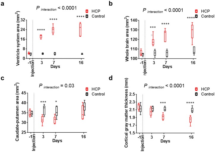
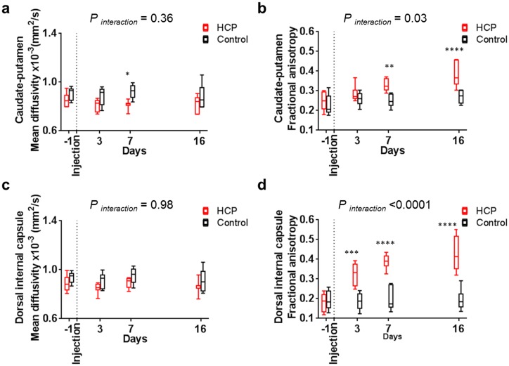
Understanding neural injury in hydrocephalus and how the brain changes during the course of the disease in-vivo remain unclear. This study describes brain deformation, microstructural and mechanical properties changes during obstructive hydrocephalus development in a rat model using multimodal magnetic resonance (MR) imaging. Hydrocephalus was induced in eight Sprague-Dawley rats (4 weeks old) by injecting a kaolin suspension into the cisterna magna. Six sham-injected rats were used as controls. MR imaging (9.4T, Bruker) was performed 1 day before, and at 3, 7 and 16 days post injection. T2-weighted MR images were collected to quantify brain deformation. MR elastography was used to measure brain stiffness, and diffusion tensor imaging (DTI) was conducted to observe brain tissue microstructure. Results showed that the enlargement of the ventricular system was associated with a decrease in the cortical gray matter thickness and caudate-putamen cross-sectional area (P < 0.001, for both), an alteration of the corpus callosum and periventricular white matter microstructure (CC+PVWM) and rearrangement of the cortical gray matter microstructure (P < 0.001, for both), while compression without gross microstructural alteration was evident in the caudate-putamen and ventral internal capsule (P < 0.001, for both). During hydrocephalus development, increased space between the white matter tracts was observed in the CC+PVWM (P < 0.001), while a decrease in space was observed for the ventral internal capsule (P < 0.001). For the cortical gray matter, an increase in extracellular tissue water was significantly associated with a decrease in tissue stiffness (P = 0.001). To conclude, this study characterizes the temporal changes in tissue microstructure, water content and stiffness in different brain regions and their association with ventricular enlargement. In summary, whilst diffusion changes were larger and statistically significant for majority of the brain regions studied, the changes in mechanical properties were modest. Moreover, the effect of ventricular enlargement is not limited to the CC+PVWM and ventral internal capsule, the extent of microstructural changes vary between brain regions, and there is regional and temporal variation in brain tissue stiffness during hydrocephalus development.


Conflict of interest statement

Competing Interests: The authors have declared that no competing interests exist.

Figures

Fig 1
Fig 1. Brain deformation variables.
Brain deformation was assessed during hydrocephalus development by quantifying changes in cross-sectional areas of the ventricular system (pink ROI, a), the whole brain (blue ROI, b), the caudate-putamen as the sum of the right and left caudate-putamen (green ROIs, c), and the thickness of the cortical gray matter as measured from the pial surface to the subcortical white matter (averaged over 5 locations, d) from the MR anatomical images.
Fig 2
Fig 2. Typical diffusion and shear modulus maps of an hydrocephalic rat brain obtained with a 9.4 T (Bruker).
(a) Mean diffusivity map (× 10−3 mm2/s). (b) Fractional anisotropy map. (c) Colored fractional anisotropy with seven ROIs: 1- corpus callosum and periventricular white matter (CC+PVWM, i.e area of the corpus callosum extending all the way to the dorsolateral angle of the lateral ventricle, violet), 2- external and 3- ventral internal capsule (pink and yellow respectively), 4- cortical gray matter (i.e area over the corpus callosum and the roof the lateral ventricles from the periventricular white matter to the pial surface, golden), 5- upper cortical gray matter (i.e top layers of the cortical gray matter excluding edematous region, red), 6- caudate-putamen (i.e average over left and right caudate-putamen, green), 7- dorsal internal capsule (turquoise). (d) Shear modulus (G*) map at 800 Hz (kPa).
Fig 3
Fig 3. Brain deformation during hydrocephalus development.
Brain deformation of hydrocephalic rats () were characterized from day three by a significantly (a) higher ventricle system cross-sectional area and (b) whole brain cross-sectional area, (c) a smaller caudate-putamen cross-sectional area at day three and day seven and also (d) a thinner cortical gray matter from day seven than control rats (□). P values for significance of the interaction between groups in the RM two-way ANOVA tests and significant Sidak’s comparisons are reported for each graph.
Fig 4
Fig 4. Typical T2-weighted anatomical MR images of rat brains obtained one day prior and three, seven, and sixteen days post kaolin injection (upper row) and sham injection (lower row).
From day three, a hyperintense signal (indicated by a white arrow) covering the corpus callosum, periventricular white matter, the external capsule and the deep regions of the cortical gray matter was observed in hydrocephalic brains but not in controls, as seen on the magnified panels. This hyperintense signal was attributed to an increase in water content of the tissue. Note: The brightness of all images has been increased by 20% to improve the visibility for readers. Abbreviations: periventricular white matter (PVWM), corpus callosum (cc), external capsule (ec), and lateral ventricles (LV).
Fig 5
Fig 5. Changes in diffusion properties in the corpus callosum + periventricular white matter (CC+PVWM), ventral internal capsule and external capsule during hydrocephalus development.
Diffusion properties in hydrocephalic rats () in the CC+PVWM (a) a higher mean diffusivity and (b) lower fractional anisotropy than for controls (□) from day three. In the ventral internal capsule, (c) the mean diffusivity was lower from day seven and (d) the fractional anisotropy was higher than for controls (□) from day three. P values for significance of the interaction between groups in the RM two-way ANOVA tests and significant Sidak’s comparisons are reported for each graph.
Fig 6
Fig 6. Changes in diffusion properties in cortical gray matter during hydrocephalus development.
Diffusion properties of the cortical gray matter of hydrocephalic rats () had a (a) higher mean diffusivity than controls (□) at day sixteen and (b) lower fractional anisotropy than controls (□) from day seven. The upper cortical gray matter region in the presence of enlarged ventricles had (c) the same mean diffusivity as controls but a (d) lower fractional anisotropy from day three. P values for significance of the interaction between groups in the RM ANOVA tests and significant Sidak’s comparisons are reported for each graph.
Fig 7
Fig 7. Changes in diffusion properties in the caudate-putamen during hydrocephalus development.
Diffusion properties showed that the mean diffusivity of the (a) caudate-putamen and (c) dorsal internal capsule was similar in hydrocephalic rats () and controls (□). On the other hand, (b) the fractional anisotropy in caudate-putamen was significantly higher from day seven and (d) the dorsal internal capsule was significantly higher from day three in hydrocephalic rats () and controls (□). P values for significance of the interaction between groups in the RM ANOVA tests and significant Sidak’s comparisons are reported for each graph.
Fig 8
Fig 8. Changes in mechanical properties in the cortical gray matter and caudate-putamen during hydrocephalus development.
Mechanical properties of the (a) cortical gray matter in hydrocephalic rats () showed a non-significant trend towards increased stiffness at day three and then decreased significantly by day seven, not seen in controls (□). In contrast, (b) the caudate-putamen stiffness in hydrocephalic rats was significantly higher at day three and then non-significant trended towards decreased stiffness. Significant t-tests are reported for each graph.
Fig 9
Fig 9. Typical histological sections stained with luxol fast blue and cresyl violet obtained from hydrocephalic and control brains sixteen days after kaolin / sham injection.
In the hydrocephalic rats, macroscopic observations (top) showed a thinner cortical gray matter which seemed to have lost some of its ground surface in the area of the cingulum and a flattened caudate-putamen. In addition, on the higher magnitude panels (bottom), in hydrocephalic brain results showed the space between tracts increased in the corpus callosum and periventricular white matter. This was not seen in matching regions in controls. This was also not observed in the ventral internal capsule of hydrocephalic (c) and control rats. Finally, only in hydrocephalic rats, the ependymal cells lining the internal surface of the enlarged ventricles were flattened and their arrangement was discontinuous. Abbreviations: ventral internal capsule (VIC), periventricular white matter (PVWM).

Similar articles

Cited by

References

    1. Williams MA, McAllister JP, Walker ML, Kranz DA, Bergsneider M, Del Bigio MR, et al. Priorities for hydrocephalus research: report from a National Institutes of Health-sponsored workshop. Journal of Neurosurgery. 2007;107(5):345–57. 10.3171/PED-07/11/345 - DOI - PubMed
    1. Del Bigio MR. Neuropathological changes caused by hydrocephalus. Acta Neuropathol. 1993;85(6):573–85. - PubMed
    1. Wills KE. Neuropsychological Functioning in Children with Spina-Bifida and or Hydrocephalus. J Clin Child Psychol. 1993;22(2):247–65.
    1. Hanigan WC, Morgan AM, Anderson RJ, Bradle P, Cohen HS, Cusack TJ, et al. Incidence and neurodevelopmental outcome of periventricular hemorrhage and hydrocephalus in a regional population of very low birth weight infants. Neurosurgery. 1991;29(5):701–6. - PubMed
    1. Vinchon M, Rekate H, Kulkarni AV. Pediatric hydrocephalus outcomes: a review. Fluids Barriers CNS. 2012;9(1):18 10.1186/2045-8118-9-18 - DOI - PMC - PubMed

Publication types