. 2016 Feb 5;11(2):e0148353.
doi: 10.1371/journal.pone.0148353. eCollection 2016.

Fluorescence In Vivo Hybridization (FIVH) for Detection of Helicobacter pylori Infection in a C57BL/6 Mouse Model

Affiliations

Fluorescence In Vivo Hybridization (FIVH) for Detection of Helicobacter pylori Infection in a C57BL/6 Mouse Model

Sílvia Fontenete et al. PLoS One. .

Abstract

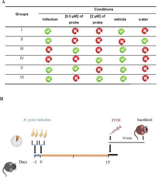
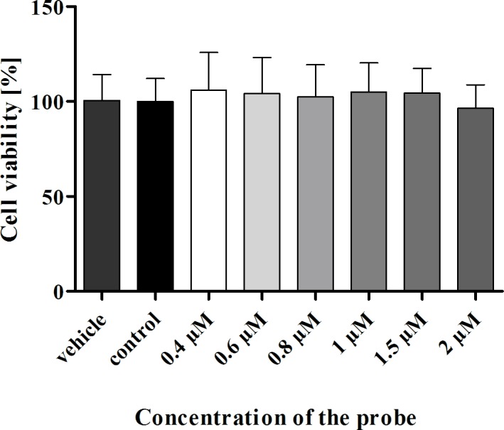
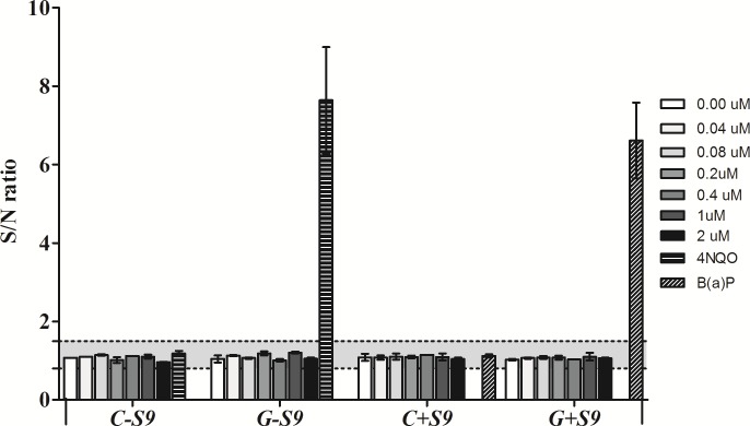
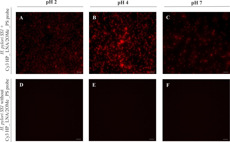
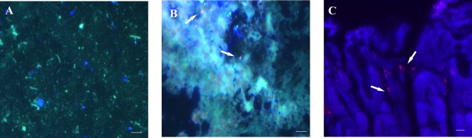
Introduction: In this study, we applied fluorescence in vivo hybridization (FIVH) using locked nucleic acid (LNA) probes targeting the bacterial rRNA gene for in vivo detection of H. pylori infecting the C57BL/6 mouse model. A previously designed Cy3_HP_LNA/2OMe_PS probe, complementary to a sequence of the H. pylori 16S rRNA gene, was used. First, the potential cytotoxicity and genotoxicity of the probe was assessed by commercial assays. Further, the performance of the probe for detecting H. pylori at different pH conditions was tested in vitro, using fluorescence in situ hybridization (FISH). Finally, the efficiency of FIVH to detect H. pylori SS1 strain in C57BL/6 infected mice was evaluated ex vivo in mucus samples, in cryosections and paraffin-embedded sections by epifluorescence and confocal microscopy.

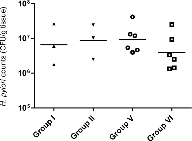
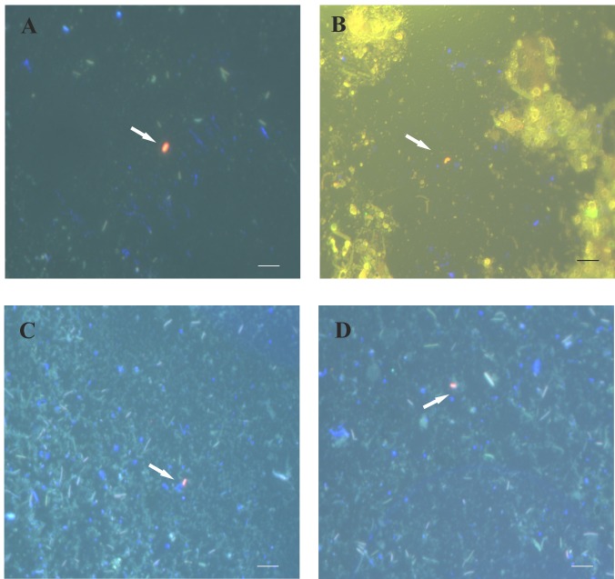
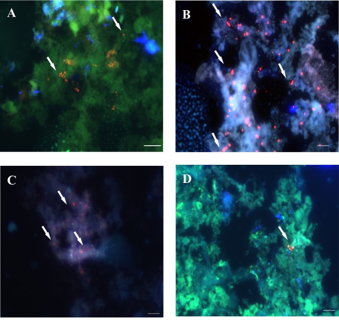
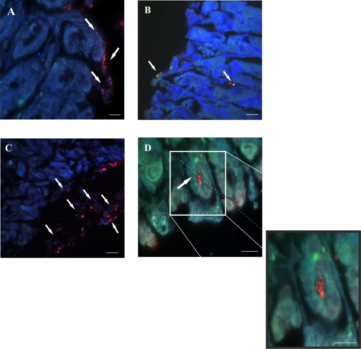
Results: H. pylori SS1 strain infecting C57BL/6 mice was successfully detected by the Cy3_HP_LNA/2OMe_PS probe in the mucus, attached to gastric epithelial cells and colonizing the gastric pits. The specificity of the probe for H. pylori was confirmed by microscopy.

Conclusions: In the future this methodology can be used in combination with a confocal laser endomicroscope for in vivo diagnosis of H. pylori infection using fluorescent LNA probes, which would be helpful to obtain an immediate diagnosis. Our results proved for the first time that FIVH method is applicable inside the body of a higher-order animal.


Conflict of interest statement

Competing Interests: The authors have read the journal's policy and the authors of this manuscript have the following competing interests: Two of the authors of this manuscript have commercial interests in this area. Jesper Wengel is cofounder of RiboTask ApS which offer LNA/2’-OMe-RNA probes for RNA targeting. Nuno Filipe Azevedo is cofounder of Biomode SA which develops molecular methods for the rapid detection of microorganisms. This does not alter the authors' adherence to PLOS ONE policies on sharing data and materials.

Figures

Fig 1
Fig 1. FIVH scheme used for detecting H. pylori in C57BL/6 mice.
A. Four control groups with n = 3 (group I to IV) were used to evaluate the background of the vehicle (group I) and tissue (group II) and the specificity of the Cy3_HyP_LNA/2’OMe probe (groups III and IV). Two tests groups (n = 6, each group) were used with different probe concentrations, 0.5 μM (group V) and 2 μM (group VII). B. Animal study protocol depicting H. pylori inoculation, time of infection in C57BL/6 mice, and FIVH after 15 days post-infection.
Fig 2
Fig 2. Effect of the Cy3_HP_LNA/2OMe_PS probe on viability of AGS gastric epithelial cells using the MTS assay.
AGS cells were treated with a range of concentrations of the Cy3_HP_LNA/2OMe_PS probe for 24 h (0.4 μM to 2 μM). Results are expressed as the mean ± SEM of three independent experiments, performed in triplicate; No statistical significant differences were found regarding probe-treated vs untreated control cells (p>0.05 by ANOVA).
Fig 3
Fig 3. VITOTOX® assay for detection of signs of genotoxicity caused by the Cy3_HyP_LNA/2’OMe probe.
Results are expressed as the signal-to-noise (S/N) ratio between exposed and unexposed VITOTOX® test bacteria (Genox or Cytox strain) in the absence or presence of S9 mix. Bap:benzo[α]pyrene, the positive control, only turns genotoxic after S9 metabolisation.
Fig 4
Fig 4. Detection of H. pylori SS1 in slides, by fluorescence in situ hybridization (FISH) using the Cy3_HP_LNA/2OMe_PS probe at different pH values, analyzed by epifluorescence microscopy.
A to C—FISH protocol performed on smears of pure cultures of H. pylori SS1 incubated with 0.2 μM of probe. D to F—FISH protocol on smears of H. pylori SS1 cultures without probe used as negative control. A and D—Experiments performed at pH 2. B and E—Experiments performed at pH 4. C and F—Experiments performed at pH 7. All images were taken at equal exposure times. Scale bar: 10 μm.
Fig 5
Fig 5. Viable bacterial counts (CFU, colony-forming units) from stomachs of mice infected with H. pylori SS1 strain for 2 weeks.
Scatter plot of CFU for each animal, with bars representing the mean value.
Fig 6
Fig 6. Detection of H. pylori SS1 in the surface of gastric mucus from mice subjected to FIVH with 0.5 μM (A and B; group V) and 2 μM (C and D; group VI) of the Cy3_HP_LNA/2OMe_PS probe.
Samples were collected and visualized directly using the epifluorescence microscope. Arrows indicate free-swimming H. pylori in the surface of mice gastric mucus. All images were taken at equal exposure times and are representative of the respective test group. Channels red, green and DAPI are overlapped. Scale bars: 10 μm.
Fig 7
Fig 7. Detection of H. pylori SS1 in frozen sections of gastric mucosa from mice subjected to FIVH, 30 min before euthanasia.
A and B: mice from group V (0.5 μM of the Cy3_HP_LNA/2OMe_PS probe), C and D: group VI (2 μM of the Cy3_HP_LNA/2OMe _PS probe). Arrows indicate the presence of H. pylori infecting the gastric mucosa. All images were taken at equal exposure times, with overlapping of the red, green and DAPI channels. All the images from each group are representative of n = 6 mice. A and C- scale bars: 50 μm. B and D- scale bars: 10 μm.
Fig 8
Fig 8. Detection of H. pylori SS1 in paraffin sections of gastric mucosa from mice test groups subjected to FIVH with the Cy3_HP_LNA/2OMe_PS probe 30 min before being sacrificed.
A and C. Fluorescence images of the detection in the surface of the gastric mucosa. B and D. Fluorescence images of the detection in the epithelium. A-B images obtained from mouse stomachs from group V. C-D images obtained from mouse stomachs from group VI. Red, green and DAPI channels are overlapped. All the images from each group are representative of n = 6 mice. A, B and C scale bars: 10 μm; D scale bars: 50 μm.
Fig 9
Fig 9. Retention of the Cy3_HP_LNA/2OMe_PS probe in the mouse stomach infected with H. pylori SS1 for 5 days, and subjected to a FIVH period of 24 hrs before being sacrificed.
A. Sample of gastric mucus. B. Frozen section of mouse gastric mucosa. C. Paraffin section of mouse gastric mucosa. Detection of H. pylori SS1 by the Cy3-labeled probe is depicted by white arrows. A and B: Red, green and DAPI channels were overlapped. C. Red and DAPI channels are overlapped. All the images from each group are representative of n = 3 mice. Scale bars: 10 μm.

Similar articles

Cited by

References

    1. Peek RM Jr., Blaser MJ (2002) Helicobacter pylori and gastrointestinal tract adenocarcinomas. Nat Rev Cancer 2: 28–37. - PubMed
    1. Milne AN, Carneiro F, O'Morain C, Offerhaus GJ (2009) Nature meets nurture: molecular genetics of gastric cancer. Hum Genet 126: 615–628. 10.1007/s00439-009-0722-x - DOI - PMC - PubMed
    1. Testerman TL, Morris J (2014) Beyond the stomach: an updated view of Helicobacter pylori pathogenesis, diagnosis, and treatment. World J Gastroenterol 20: 12781–12808. 10.3748/wjg.v20.i36.12781 - DOI - PMC - PubMed
    1. Lopes AI, Vale FF, Oleastro M (2014) Helicobacter pylori infection—recent developments in diagnosis. World J Gastroenterol 20: 9299–9313. 10.3748/wjg.v20.i28.9299 - DOI - PMC - PubMed
    1. Gheonea DI, Cartana T, Ciurea T, Popescu C, Badarau A, Saftoiu A (2011) Confocal laser endomicroscopy and immunoendoscopy for real-time assessment of vascularization in gastrointestinal malignancies. World J Gastroenterol 17: 21–27. 10.3748/wjg.v17.i1.21 - DOI - PMC - PubMed

Publication types