. 2016 Mar 1;7(9):10090-103.
doi: 10.18632/oncotarget.7136.

CD93 and dystroglycan cooperation in human endothelial cell adhesion and migration adhesion and migration

Affiliations

CD93 and dystroglycan cooperation in human endothelial cell adhesion and migration adhesion and migration

Federico Galvagni et al. Oncotarget. .

Abstract

CD93 is a transmembrane glycoprotein predominantly expressed in endothelial cells. Although CD93 displays proangiogenic activity, its molecular function in angiogenesis still needs to be clarified. To get molecular insight into the biological role of CD93 in the endothelium, we performed proteomic analyses to examine changes in the protein profile of endothelial cells after CD93 silencing. Among differentially expressed proteins, we identified dystroglycan, a laminin-binding protein involved in angiogenesis, whose expression is increased in vascular endothelial cells within malignant tumors. Using immunofluorescence, FRET, and proximity ligation analyses, we observed a close interaction between CD93 and β-dystroglycan. Moreover, silencing experiments showed that CD93 and dystroglycan promoted endothelial cell migration and organization into capillary-like structures. CD93 proved to be phosphorylated on tyrosine 628 and 644 following cell adhesion on laminin through dystroglycan. This phosphorylation was shown to be necessary for a proper endothelial migratory phenotype. Moreover, we showed that during cell spreading phosphorylated CD93 recruited the signaling protein Cbl, which in turn was phosphorylated on tyrosine 774. Altogether, our results identify a new signaling pathway which is activated by the cooperation between CD93 and dystroglycan and involved in the control of endothelial cell function.

Keywords: C1qRp; Cbl; Src; angiogenesis; signal transduction.


Conflict of interest statement

CONFLICTS OF INTEREST

The authors declare no conflicts of interest.

Figures

Figure 1
Figure 1. In ECs DG and CD93 silencing reveals adaptive changes in expression of both proteins
(A) HUVEC were infected with lentiviral vectors expressing unrelated (unr) or CD93 shRNAs (clones 85 and 86). Total cell lysates were obtained from exponentially growing cells and subjected to comparative proteomic analysis. Expanded views from the 2-DE gels show the increased expression pattern of β-DG in CD93-silenced cells compared to cells expressing an unrelated shRNA. Arrows indicate the experimental coordinates (pI and Mr) of the spot identified as β-DG by mass spectrometry. (B) HUVEC were infected as in A. Cell extracts were analyzed by Western blotting using anti-CD93 and anti-β-DG antibodies. Anti-β-actin antibodies were used to confirm equal protein loading. Not infected cells (HUVEC). (C) Quantitative analysis of β-DG protein levels from independent experiments performed as in B. Protein levels were quantified by densitometric scanning and the values, normalized to β-actin protein levels, were averaged and expressed as arbitrary units. (D) ECs were infected with lentiviral vectors expressing unrelated (unr) or DG shRNAs (clones C7 and C10). Cell extracts were analyzed by Western blotting as in B. (E) Quantitative analysis of CD93 protein levels performed as in D, normalized to β-actin protein levels, and expressed as arbitrary units. Representative images from a triplicate set are shown and data represent the means ± SD of three independent experiments.
Figure 2
Figure 2. Direct association between CD93 and β-DG in ECs
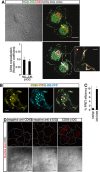
(A) HUVEC, plated onto laminin-coated glass coverslips, were fixed during the late phase of spreading and analyzed by immunofluorescence using anti-β-DG and anti-CD93 antibodies. Differential interference contrast (DIC), overlay of stained cells, and white dot colocalization images are shown. Plot shows quantification (using Manders' coefficient) of CD93 colocalization with β-DG at the cell margin (c.m.) and in whole cells (w.c.) (mean ± SD; cells = 21; n = 3). Scale bar, 12 μm. In the inset white dots show CD93 and β-DG colocalization at the cell margin. Scale bar of the inset is 3 μm. (B) CD93-YFP and β-DG-CFP were cotransfected into ECs. Fully spread cells on laminin-coated surfaces were fixed and subjected to immunofluorescence. Immunofluorescence shows CD93 and β-DG colocalization both at the plasma membrane and within intracellular vesicles. Scale bar, 8 μm. (C) Cells treated as in B were subjected to FRET analyses. The mean value of the FRET efficiency between acceptor (CD93-YFP) and donor (β-DG-CFP) was 9.11 ± 0.84%, after subtraction of the background. FRET data represent the means ± SD of three independent experiments, carried out on different days and with different cell preparations. (D) Representative confocal images of CD93/β-DG protein interaction detected in situ by Duolink stain. HUVEC exponentially growing on laminin-coated surfaces were fixed and treated at the same time with mouse anti-CD93 and rabbit anti-β-DG antibodies (CD93-β-DG). Close proximity of the primary antibodies was revealed by localized amplification. Protein-protein interactions were visualized as individual spots by red fluorescence. Background was assayed by removing one of the two primary antibodies from the reaction (anti-β-DG antibodies removed, neg. contr. CD93; anti-CD93 removed, neg. contr. β-DG). DIC images of stained cells are shown. The corresponding cell boundary is indicated by white dotted lines. Experiment was performed three times. Scale bars represent 18 μm.
Figure 3
Figure 3. CD93 or DG knockdown impairs EC function
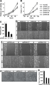
HUVEC were infected with a lentiviral vector expressing unrelated (unr), or CD93 (clones 85 or 86), or DG (clones C7 or C10) shRNAs. Not infected ECs were also analyzed (HUVEC). (A) Cell viability assay performed at the indicated time points on infected HUVEC plated in 96-well plates and grown in complete medium. The optical density values were expressed as relative cell viability. (B) ECs were infected, plated in 24-well plates and grown in complete medium. Cell number was evaluated by using a hemocytometer at the indicated time points. (C) Migration assay on infected HUVEC. Cells were grown in growth factor-depleted culture medium and plated in a Boyden chamber. Chemotaxis was stimulated with 10 ng/ml VEGF (Sigma-Aldrich). Migratory cells were stained and counted under a light microscope. (D and E) Wound healing assays of HUVEC infected as indicated. Cell monolayers were wounded with a sterile pipette tip, washed, and grown in complete medium. Cells were observed under a light microscope and photographed at 0 and 8 h. Representative experiments are shown (original magnification, x100). (F) HUVEC infected as indicated were grown in complete medium on Matrigel and the formation of capillary-like structures was observed 20 h after seeding. A representative experiment is shown (original magnification, ×100). (G) Quantification of tube length was performed based on the results shown in panel F. Results were expressed as means ± SD of four different fields randomly chosen from each group. All data represent the means ± SD of three-five independent experiments.
Figure 4
Figure 4. Binding of DG to laminin induces CD93 phosphorylation via Src
(A) The schematic diagram illustrates the domains of CD93 and the 47-amino acid sequence of its cytoplasmic tail containing tyrosine 628 and 644. CTLD, C-type lectin-like domain; EGF-like, Epidermal Growth Factor repeats; Mucin. Mucin-like domain; TM, transmembrane domain. (B) Cell extracts from ECs spreading and growing on gelatin or laminin were immunoprecipitated with anti-CD93 antibodies. Immunoprecipitates were analyzed by Western blotting with anti-phosphotyrosine and anti-CD93 antibodies to confirm equal loading. (C) HUVEC were infected with a lentiviral vector expressing unrelated (unr) or DG (clones C7 or C10) shRNAs. Not infected ECs were also analyzed (HUVEC). Cell extracts from cells spreading and growing on laminin were immunoprecipitated or not (+, −) with anti-CD93 antibodies and analyzed by Western blotting with anti-phosphotyrosine antibodies. To confirm equal loading, whole cell lysates were analyzed by Western blotting with anti-CD93 and anti-β-actin antibodies. (D) HUVEC were allowed to adhere and grow on laminin in the presence (+) or not (−) of PP2 (10 μm). Cell lysates were immunoprecipitated with anti-CD93 antibodies and analyzed by immunoblotting with anti-phosphotyrosine and anti-CD93 antibodies to confirm equal loading. In the same cell lysates, phosphorylation on tyrosine 416 of Src, a protein modification that is closely correlated with kinase activity, was analyzed by Western blotting with anti-phospho-Y416 Src and anti-Src antibodies to confirm equal loading. All experiments were performed three-four times. (E) Human Lenti-X 293T cells, which do not express wild type CD93, were transiently cotransfected with a construct expressing human CD93 and the constitutively active (DP) or kinase dead (DN) Src kinase. Transfection with an empty vector (mock) is indicated. Cell lysates were immunoprecipitated with anti-CD93 antibodies and analyzed by Western blotting with anti-phosphotyrosine and anti-CD93 antibodies to confirm equal loading.
Figure 5
Figure 5. CD93 phosphorylation stimulates EC migration
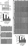
HUVEC were infected with lentiviral particles expressing CD93 shRNA clone 85 alone (85) or in combination (85+) with each CD93 mutant (res85, CD93 resistant to silencing; res85Y1, CD93 resistant to silencing mutated in tyrosine 628; res85Y2, CD93 resistant to silencing mutated in tyrosine 644; res85Y1Y2, CD93 resistant to silencing mutated in tyrosine 628 and 644). Control cells were infected with an unrelated shRNA (unr). (A) Cell extracts from infected ECs were analyzed by immunoblotting using anti-CD93, anti-β-DG, and anti-β-actin antibodies to confirm equal loading. (B) Wound healing assay of HUVEC infected as indicated. Cell monolayers were wounded with a sterile pipette tip, washed, and grown in complete medium. Cells were observed under a light microscope and photographed at 0 and 16 h. A representative experiment is shown (original magnification, ×100). (C) Migration assay on infected HUVEC. Cells were grown in growth factor-depleted culture medium and plated in a Boyden chamber. Chemotaxis was stimulated with 10 ng/ml VEGF. Migratory cells were stained and counted under a light microscope. (D) ECs infected as indicated were grown in complete medium on Matrigel and the formation of vascular capillary networks was observed 20 h after seeding. A representative experiment is shown (original magnification, ×100). (E) Quantification of tube length was performed based on the results shown in panel D. Results were expressed as means ± SD of four different fields randomly chosen from each group. All data represent the means ± SD of three independent experiments.
Figure 6
Figure 6. During EC spreading, Cbl is recruited to CD93 and phosphorylated on tyrosine 774
(A) Sequence alignment of Cbl recruitment sites previously characterized in different signaling proteins. Colored as opposed to gray residues are generally conserved. Green residues are hydrophobic, blue residues basic, and violet residues polar. Three dots indicate that the protein sequence continues. EGFR, epithelial growth factor receptor; NTR, neurotrophin receptor; VEGFR, vascular endothelial growth factor receptor; APS, adapter with pleckstrin homology and SH2 domains; Lnk, lymphocyte adaptor protein. APS, Lnk, and SH2-B belong to a family of adapter proteins that are implicated in signaling transduction [26]. (B) HUVEC were released from culture plates by EDTA treatment, plated on laminin-coated surface in complete medium, and allowed to spread. Cell lysates from spreading cells were immunoprecipitated with and without (No Ab) anti-CD93 antibodies. Immunoprecipitates were analyzed by Western blotting with anti-Cbl antibodies. Whole cell extracts were used to check the molecular size of the coimmunoprecipitated proteins (Cell ex.). Equal input was confirmed by Western blotting using anti-β-actin antibodies. (C) HUVEC were infected with a lentiviral vector expressing unrelated (unr) or DG (clones C7 or C10) shRNAs. Cell lysates were immunoprecipitated with anti-CD93 antibodies and analyzed by Western blotting with anti-Cbl and anti-CD93 antibodies to confirm equal loading. (D) Human Lenti-X 293T cells do not express wild type CD93 [5]. Cells were transiently cotransfected with constructs expressing CD93 mutants (res85, res85Y1, res85Y2, and res85Y1Y2) and the constitutively active (dominant positive, DP) or kinase dead (dominant negative, DN) Src kinase. Cell extracts were immunoprecipitated with anti-CD93 antibodies and analyzed by Western blotting with anti-Cbl, anti-phosphotyrosine, and anti-CD93 antibodies to confirm equal loading. In the presence of active Src, only wild type CD93 (res85) displays a phosphorylation signal and interacts with Cbl. (E) ECs were detached from culture plates and replated on laminin-coated surfaces. Cell extracts obtained at different degrees of cell spreading were analyzed by Western blotting using anti-phospho-Cbl(Y774) antibodies. 15′ and 2 h indicate early and late spreading cells respectively. 24 h after replating, cells reached confluency. (F) ECs were fixed during the spreading phases and analyzed by immunofluorescence using anti-CD93 and anti-phospho-Cbl(Y774) antibodies. Overlay of stained cells and white dot colocalization images are shown. Plot shows quantification (using Manders' coefficient) of CD93 colocalization with phospho-Cbl(Y774) at the cell margin (c.m.) and in whole cells (w.c.) (mean ± SD; cells = 20; n = 3). Scale bar, 14 μm. In the inset white dots show CD93 and phospho-Cbl(Y774) colocalization at the cell margin. Scale bar of the inset is 3 μm. (G) HUVEC were infected with lentiviral vectors expressing unrelated (unr) or CD93 (clones 85 or 86) shRNAs. Lysates obtained from spreading cells were analyzed by Western blotting using anti-phospho-Cbl(Y774) antibodies. In E and G to confirm equal protein loading, whole cell lysates were analyzed by Western blotting with anti-Cbl and anti-β-actin antibodies. Every experiment was repeated at least three times.

Similar articles

Cited by

References

    1. Lamalice L, Le Boeuf F, Huot J. Endothelial cell migration during angiogenesis. Circ Res. 2007;100:782–794. - PubMed
    1. Carmeliet P. Angiogenesis in health and disease. Nat Med. 2003;9:653–660. - PubMed
    1. Schmidt S, Friedl P. Interstitial cell migration: integrin-dependent and alternative adhesion mechanisms. Cell Tissue Res. 2010;339:83–92. - PMC - PubMed
    1. Greenlee MC, Sullivan SA, Bohlson SS. CD93 and related family members: their role in innate immunity. Curr Drug Targets. 2008;9:130–138. - PubMed
    1. Orlandini M, Galvagni F, Bardelli M, Rocchigiani M, Lentucci C, Anselmi F, Zippo A, Bini L, Oliviero S. The characterization of a novel monoclonal antibody against CD93 unveils a new antiangiogenic target. Oncotarget. 2014;5:2750–2760. doi: 10.18632/oncotarget.1887. - DOI - PMC - PubMed

Publication types

MeSH terms