

A humanized antibody for imaging immune checkpoint ligand PD-L1 expression in tumors
- PMID: 26848870
- PMCID: PMC4891115
- DOI: 10.18632/oncotarget.7143
A humanized antibody for imaging immune checkpoint ligand PD-L1 expression in tumors
Abstract
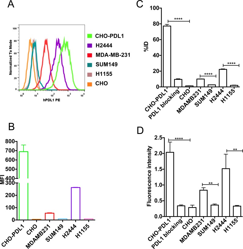
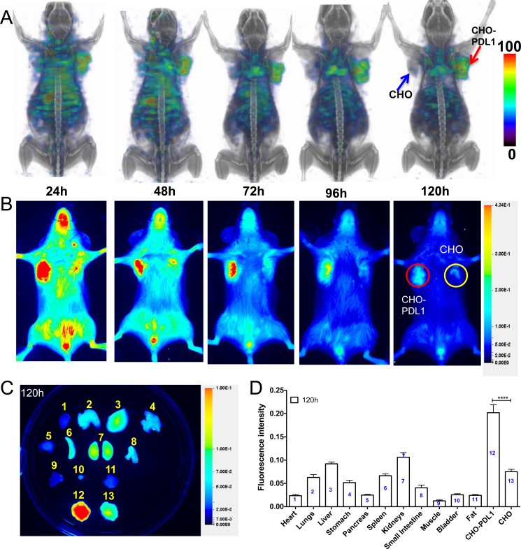
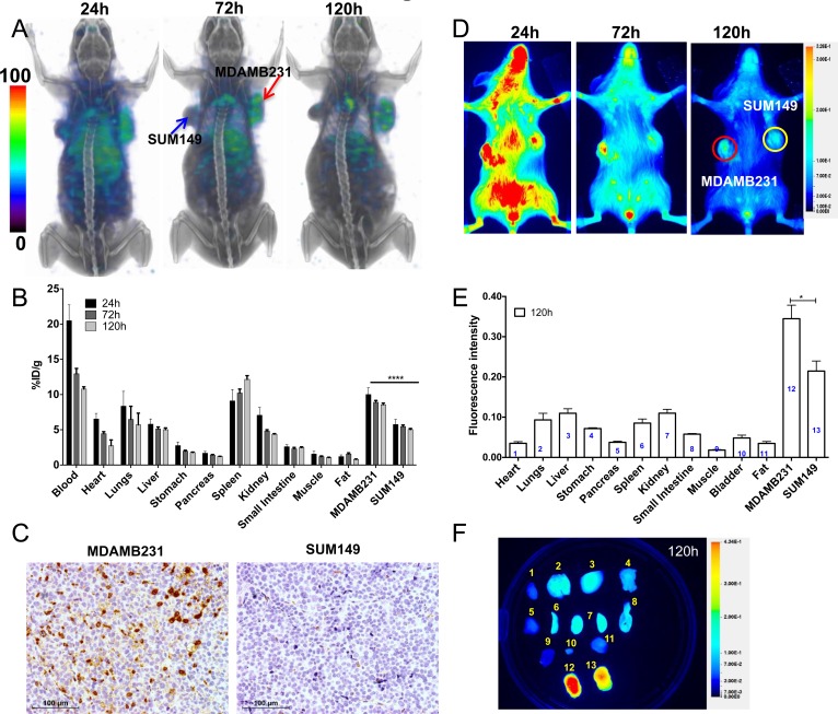
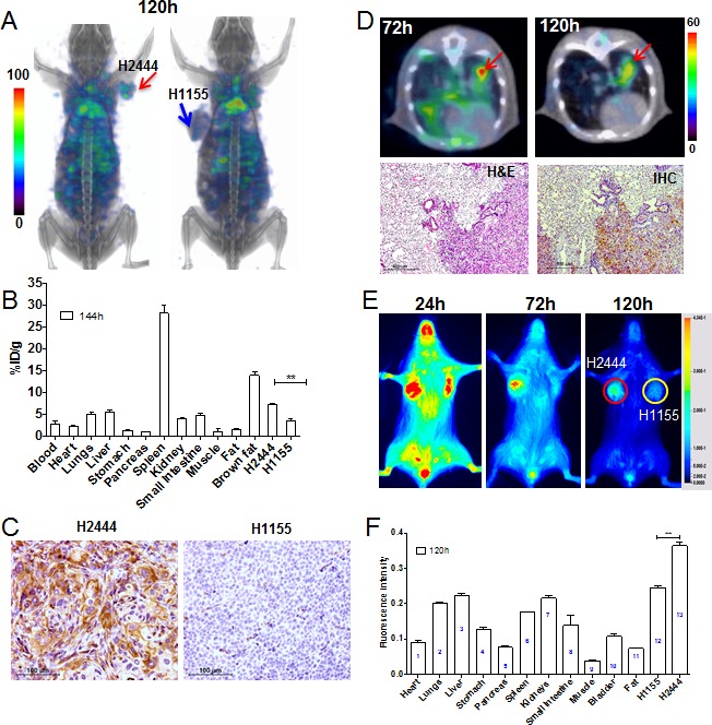
Antibodies targeting the PD-1/PD-L1 immune checkpoint lead to tumor regression and improved survival in several cancers. PD-L1 expression in tumors may be predictive of response to checkpoint blockade therapy. Because tissue samples might not always be available to guide therapy, we developed and evaluated a humanized antibody for non-invasive imaging of PD-L1 expression in tumors. Radiolabeled [111In]PD-L1-mAb and near-infrared dye conjugated NIR-PD-L1-mAb imaging agents were developed using the mouse and human cross-reactive PD-L1 antibody MPDL3280A. We tested specificity of [111In]PD-L1-mAb and NIR-PD-L1-mAb in cell lines and in tumors with varying levels of PD-L1 expression. We performed SPECT/CT imaging, biodistribution and blocking studies in NSG mice bearing tumors with constitutive PD-L1 expression (CHO-PDL1) and in controls (CHO). Results were confirmed in triple negative breast cancer (TNBC) (MDAMB231 and SUM149) and non-small cell lung cancer (NSCLC) (H2444 and H1155) xenografts with varying levels of PD-L1 expression. There was specific binding of [111In]PD-L1-mAb and NIR-PD-L1-mAb to tumor cells in vitro, correlating with PD-L1 expression levels. In mice bearing subcutaneous and orthotopic tumors, there was specific and persistent high accumulation of signal intensity in PD-L1 positive tumors (CHO-PDL1, MDAMB231, H2444) but not in controls. These results demonstrate that [111In]PD-L1-mAb and NIR-PD-L1-mAb can detect graded levels of PD-L1 expression in human tumor xenografts in vivo. As a humanized antibody, these findings suggest clinical translation of radiolabeled versions of MPDL3280A for imaging. Specificity of NIR-PD-L1-mAb indicates the potential for optical imaging of PD-L1 expression in tumors in relevant pre-clinical as well as clinical settings.
Keywords: MPDL3280A; immune escape; immunotherapy; molecular imaging; personalized medicine.
Conflict of interest statement
None.
Figures
Comment on
-
Noninvasive Imaging of Immune Checkpoint Ligand PD-L1 in Tumors and Metastases for Guiding Immunotherapy.Mol Imaging. 2017 Jan-Dec;16:1536012117718459. doi: 10.1177/1536012117718459. Mol Imaging. 2017. PMID: 28707500 Free PMC article. Review.
Similar articles
-
PD-L1 Detection in Tumors Using [(64)Cu]Atezolizumab with PET.Bioconjug Chem. 2016 Sep 21;27(9):2103-10. doi: 10.1021/acs.bioconjchem.6b00348. Epub 2016 Aug 9. Bioconjug Chem. 2016. PMID: 27458027 Free PMC article.
-
Near-infrared fluorescence-labeled anti-PD-L1-mAb for tumor imaging in human colorectal cancer xenografted mice.J Cell Biochem. 2019 Jun;120(6):10239-10247. doi: 10.1002/jcb.28308. Epub 2019 Jan 4. J Cell Biochem. 2019. PMID: 30609118 Free PMC article.
-
Radioimmunoimaging and targeting treatment in an immunocompetent murine model of triple-negative breast cancer using radiolabeled anti-programmed death-ligand 1 monoclonal antibody.J Labelled Comp Radiopharm. 2018 Sep;61(11):826-836. doi: 10.1002/jlcr.3650. Epub 2018 Jul 4. J Labelled Comp Radiopharm. 2018. PMID: 29923634
-
[Efficacy of PD-1/PD-L1 immune checkpoint inhibitors and PD-L1 testing in thoracic cancers].Ann Pathol. 2017 Feb;37(1):61-78. doi: 10.1016/j.annpat.2016.12.009. Epub 2017 Feb 3. Ann Pathol. 2017. PMID: 28162296 Review. French.
-
Anti-PD-1/PD-L1 Therapy for Non-Small-Cell Lung Cancer: Toward Personalized Medicine and Combination Strategies.J Immunol Res. 2018 Aug 8;2018:6984948. doi: 10.1155/2018/6984948. eCollection 2018. J Immunol Res. 2018. PMID: 30159341 Free PMC article. Review.
Cited by
-
Sensitive and quantitative in vivo analysis of PD-L1 using magnetic particle imaging and imaging-guided immunotherapy.Eur J Nucl Med Mol Imaging. 2023 Apr;50(5):1291-1305. doi: 10.1007/s00259-022-06083-2. Epub 2022 Dec 12. Eur J Nucl Med Mol Imaging. 2023. PMID: 36504279
-
A Preclinical Assessment of 89Zr-atezolizumab Identifies a Requirement for Carrier Added Formulations Not Observed with 89Zr-C4.Bioconjug Chem. 2018 Oct 17;29(10):3476-3482. doi: 10.1021/acs.bioconjchem.8b00632. Epub 2018 Sep 24. Bioconjug Chem. 2018. PMID: 30227708 Free PMC article.
-
The combination therapy with EpCAM/CD3 BsAb and MUC-1/CD3 BsAb elicited antitumor immunity by T-cell adoptive immunotherapy in lung cancer.Int J Med Sci. 2021 Jul 31;18(15):3380-3388. doi: 10.7150/ijms.61681. eCollection 2021. Int J Med Sci. 2021. PMID: 34522164 Free PMC article.
-
Nuclear Molecular Imaging Strategies in Immune Checkpoint Inhibitor Therapy.Diagnostics (Basel). 2017 Apr 21;7(2):23. doi: 10.3390/diagnostics7020023. Diagnostics (Basel). 2017. PMID: 28430133 Free PMC article. Review.
-
Preclinical Pharmacokinetics and Dosimetry of an 89Zr Labelled Anti-PDL1 in an Orthotopic Lung Cancer Murine Model.Front Med (Lausanne). 2022 Jan 31;8:741855. doi: 10.3389/fmed.2021.741855. eCollection 2021. Front Med (Lausanne). 2022. PMID: 35174180 Free PMC article.
References
-
- Topalian SL, Sznol M, McDermott DF, Kluger HM, Carvajal RD, Sharfman WH, Brahmer JR, Lawrence DP, Atkins MB, Powderly JD, Leming PD, Lipson EJ, Puzanov I, Smith DC, Taube JM, Wigginton JM, et al. Survival, durable tumor remission, and long-term safety in patients with advanced melanoma receiving nivolumab. J Clin Oncol. 2014;32:1020–1030. - PMC - PubMed
-
- Gettinger SN, Horn L, Gandhi L, Spigel DR, Antonia SJ, Rizvi NA, Powderly JD, Heist RS, Carvajal RD, Jackman DM, Sequist LV, Smith DC, Leming P, Carbone DP, Pinder-Schenck MC, Topalian SL, et al. Overall Survival and Long-Term Safety of Nivolumab (Anti-Programmed Death 1 Antibody, BMS-936558, ONO-4538) in Patients With Previously Treated Advanced Non-Small-Cell Lung Cancer. J Clin Oncol. 2015;33:2004–2012. - PMC - PubMed
Publication types
MeSH terms
Substances
Grants and funding
LinkOut - more resources
Full Text Sources
Other Literature Sources
Medical
Research Materials
Miscellaneous
