. 2016 Feb 23;55(7):1107-19.
doi: 10.1021/acs.biochem.5b00993. Epub 2016 Feb 5.

Structural Insights into Mycobacterium tuberculosis Rv2671 Protein as a Dihydrofolate Reductase Functional Analogue Contributing to para-Aminosalicylic Acid Resistance

Affiliations

Structural Insights into Mycobacterium tuberculosis Rv2671 Protein as a Dihydrofolate Reductase Functional Analogue Contributing to para-Aminosalicylic Acid Resistance

Yu-Shan Cheng et al. Biochemistry. .

Abstract

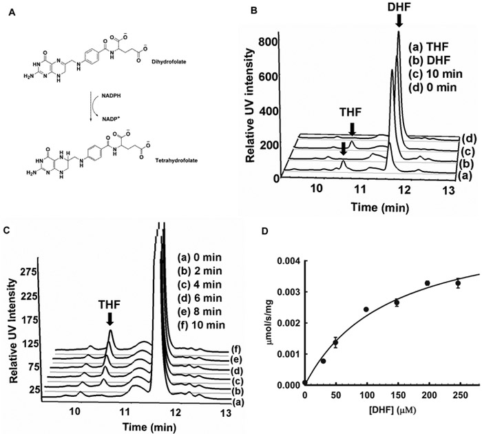
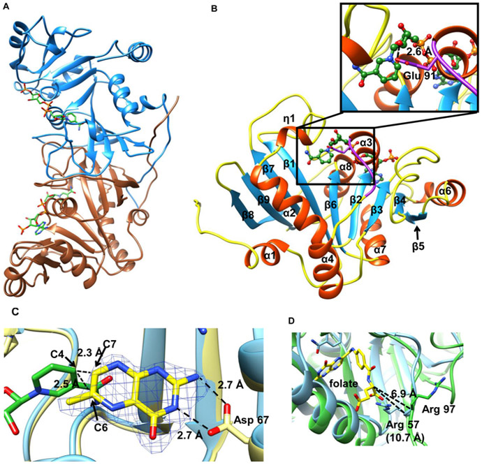
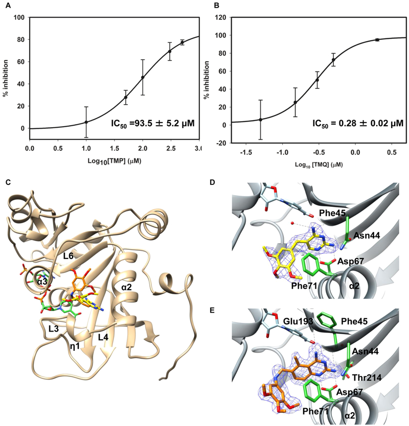
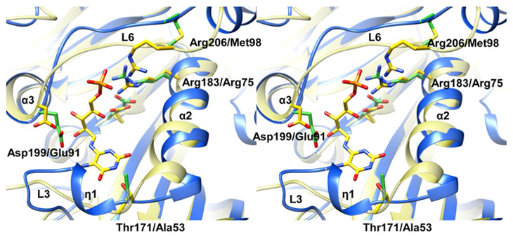
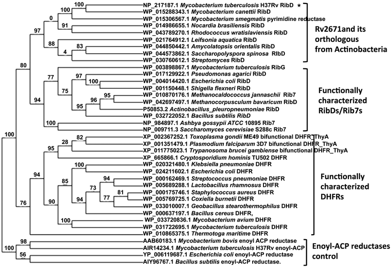
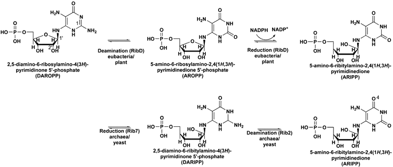
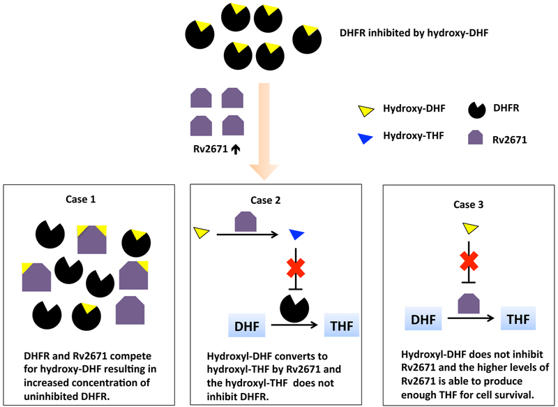
Mycobacterium tuberculosis (Mtb) Rv2671 is annotated as a 5-amino-6-ribitylamino-2,4(1H,3H)-pyrimidinedione 5'-phosphate (AROPP) reductase (RibD) in the riboflavin biosynthetic pathway. Recently, a strain of Mtb with a mutation in the 5' untranslated region of Rv2671, which resulted in its overexpression, was found to be resistant to dihydrofolate reductase (DHFR) inhibitors including the anti-Mtb drug para-aminosalicylic acid (PAS). In this study, a biochemical analysis of Rv2671 showed that it was able to catalyze the reduction of dihydrofolate (DHF) to tetrahydrofolate (THF), which explained why the overexpression of Rv2671 was sufficient to confer PAS resistance. We solved the structure of Rv2671 in complex with the NADP(+) and tetrahydrofolate (THF), which revealed the structural basis for the DHFR activity. The structures of Rv2671 complexed with two DHFR inhibitors, trimethoprim and trimetrexate, provided additional details of the substrate binding pocket and elucidated the differences between their inhibitory activities. Finally, Rv2671 was unable to catalyze the reduction of AROPP, which indicated that Rv2671 and its closely related orthologues are not involved in riboflavin biosynthesis.


Figures

Figure 1.
Figure 1.. Characterization of Rv2671 as a DHFR.
(A) Scheme of the DHF reduction reaction. (B) HPLC analysis of the in vitro reaction and the relevant control. (C) Time course for the reaction shows that the amount of THF increases with time. (D) Steady state kinetic reaction measured in the presence of Rv2671 and saturated NADPH. Reaction velocity plotted versus DHF concentration (Km = 136 ± 32 μM; kcat = 0.15 ± 0.016 s−1). Each DHF concentration was performed in triplicate, and the data represent mean values ± SD.
Figure 2.
Figure 2.. Crystal structure of Rv2671 and the THF binding mode.
(A) Ribbon representation of the dimeric Rv2671. (B) Ribbon representation of the Rv2671 in complex with NADP+. The secondary structural elements are labeled sequentially. The loop (residue 91–95) disordered in the native purified structure is shown in purple, and NADP+ is shown in the green ball and stick model. The hydrogen bond interaction between the ribose group of NADP+ and Glu 91 in the loop (residue 91–95) is shown in the zoomed-in figure. (C) Superimposition of the THF bound Rv2671 structure (yellow) with the NADP+ bound Rv2671 structure (blue). NADP+ and the pteridine ring of THF are colored in green and yellow. The simulated annealing omit map contoured at 2.5σ around the pteridine ring is shown in blue. Distances between the C4 of nicotinamide and the C6 and C7 of the pteridine are labeled. (D) Superimposition of the NADP+ bound Rv2671 structure (green) with the folate-NADPH bound E. coli RibD (4pdj, blue). Folate is colored in yellow. The side chain of the Arg 97 in the Rv2671 complex structure points to the other direction against the folate. The distances between α-carbon of the glutamate group in folate and the main chain carbon of the arginines are 6.9 and 10.7 Å, respectively.
Figure 3.
Figure 3.. Crystal structure of Rv2671 in complex with TMP or TMQ.
(A) The mean % inhibition values of Rv2671 are plotted against the log10 concentration of the DHFR inhibitor TMP. The calculated IC50 value is 93.5 ± 5.2 μM. (B) The mean % inhibition values of Rv2671 are plotted against the log10 concentration of the DHFR inhibitor TMQ. The calculated IC50 value is 0.28 ± 0.02 μM. (C) Ribbon representation of Rv2671 (tan) in complex with TMP (yellow) and TMQ (orange). (D) Binding of the TMP with Rv2671. The simulated annealing omit map contoured at 2.5σ around the inhibitor TMP is shown in blue. The key residues interacting with TMP are labeled and shown as sticks in green. The hydrogen bonds are shown as dashed lines. (E) Binding of TMQ with Rv2671. The simulated annealing omit map contoured at 2.5 σ around the inhibitor TMQ is shown in blue. The main residues interacting with TMP are labeled and shown as green sticks. Different binding modes of the trimethoxybenzyl rings of TMP and TMQ in Rv2671 can be observed in D and E. The trimethoxybenzyl ring of TMP in the complex structure is perpendicular to Phe71. The trimethoxybenzyl ring of TMQ in the complex structure is parallel to Phe71.
Figure 4.
Figure 4.
Residue differences between the substrate binding sites of Rv2671 and Bs RibG. Superposition of NADP+ bound Rv2671 (blue) with the AIF bound Bs RibG (yellow) is shown in a stereo view. AIF and the residues directly interacting with AIF in Bs RibG are shown as yellow sticks, and the corresponding residues in Rv2671 are shown as green sticks.
Figure 5.
Figure 5.
Phylogenetic tree of pyrimidine reductases and DHFRs. Sequences were aligned with MUSCLE; the tree was generated by the approximation of the standard Likelihood Ratio Test with PhyML. The numbers at the nodes indicate the level of confidence for the branches.
Scheme 1
Scheme 1
Scheme 2
Scheme 2

Similar articles

Cited by

References

    1. CDC. Update: Availability of Streptomycin and para-Amino-salicylic Acid—United States, Morbidity and Mortality Weekly Report; 41, 26; Center for Disease Control and Prevention: Atlanta, GA, 1992; 482.
    1. Chakraborty S, Gruber T, Barry CE 3rd, Boshoff HI, and Rhee KY (2013) Para-aminosalicylic acid acts as an alternative substrate of folate metabolism in Mycobacterium tuberculosis. Science, 339, 88–91. - PMC - PubMed
    1. Zheng J, Rubin EJ, Bifani P, Mathys V, Lim V, Au M, Jang J, Nam J, Dick T, Walker JR, Pethe K, and Camacho LR (2013) para-Aminosalicylic acid is a prodrug targeting dihydrofolate reductase in Mycobacterium tuberculosis. J. Biol. Chem 288, 23447–23456. - PMC - PubMed
    1. Rieckmann KH (1973) The in Vitro Activity of Experimental Antimalarial Compounds against Strains of Plasmodium falciparum with Varying Degrees of Sensitivity to Pyrimethamine and Chloroquine, in Chemotherapy of Malaria and Resistance to Antimalarials, 529th ed., p 58, WHO Tech. Rep. Ser., World Health Organization, Geneva, Switzerland.
    1. Zhang X, Liu L, Zhang Y, Dai G, Huang H, and Jin Q (2015) Genetic determinants involved in p-aminosalicylic acid resistance in clinical isolates from tuberculosis patients in northern China from 2006 to 2012. Antimicrob. Agents Chemother 59, 1320–1324. - PMC - PubMed

Publication types

MeSH terms

LinkOut - more resources