Effect of Collateral Sprouting on Donor Nerve Function After Nerve Coaptation: A Study of the Brachial Plexus
- PMID: 26848925
- PMCID: PMC4762401
- DOI: 10.12659/msm.895397
Effect of Collateral Sprouting on Donor Nerve Function After Nerve Coaptation: A Study of the Brachial Plexus
Abstract
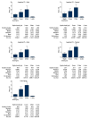
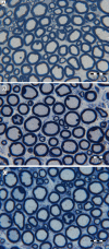
BACKGROUND The aim of the present study was to evaluate the donor nerve from the C7 spinal nerve of the rabbit brachial plexus after a coaptation procedure. Assessment was performed of avulsion of the C5 and C6 spinal nerves treated by coaptation of these nerves to the C7 spinal nerve. MATERIAL AND METHODS After nerve injury, fourteen rabbits were treated by end-to-side coaptation (ETS), and fourteen animals were treated by side-to-side coaptation (STS) on the right brachial plexus. Electrophysiological and histomorphometric analyses and the skin pinch test were used to evaluate the outcomes. RESULTS There was no statistically significant difference in the G-ratio proximal and distal to the coaptation in the ETS group, but the differences in the axon, myelin sheath and fiber diameters were statistically significant. The comparison of the ETS and STS groups distal to the coaptation with the controls demonstrated statistically significant differences in the fiber, axon, and myelin sheath diameters. With respect to the G-ratio, the ETS group exhibited no significant differences relative to the control, whereas the G-ratio in the STS group and the controls differed significantly. In the electrophysiological study, the ETS and STS groups exhibited major changes in the biceps and subscapularis muscles. CONCLUSIONS The coaptation procedure affects the histological structure of the nerve donor, but it does not translate into changes in nerve conduction or the sensory function of the limb. The donor nerve lesion in the ETS group is transient and has minimal clinical relevance.
Figures


Similar articles
-
Collateral sprouting axons of end-to-side nerve coaptation in the avulsion of ventral branches of the C5-C6 spinal nerves in the brachial plexus.Folia Neuropathol. 2015;53(4):327-42. doi: 10.5114/fn.2015.56547. Folia Neuropathol. 2015. PMID: 26785367
-
Physiologic and morphologic aspects of nerve regeneration after end-to-end or end-to-side coaptation in a rat model of brachial plexus injury.J Hand Surg Am. 2002 Jan;27(1):133-42. doi: 10.1053/jhsu.2002.30370. J Hand Surg Am. 2002. PMID: 11810627
-
The rabbit brachial plexus as a model for nerve repair surgery--histomorphometric analysis.Anat Rec (Hoboken). 2015 Feb;298(2):444-54. doi: 10.1002/ar.23058. Epub 2014 Oct 18. Anat Rec (Hoboken). 2015. PMID: 25284580
-
Chapter 14: End-to-side nerve regeneration: from the laboratory bench to clinical applications.Int Rev Neurobiol. 2009;87:281-94. doi: 10.1016/S0074-7742(09)87014-1. Int Rev Neurobiol. 2009. PMID: 19682643 Review.
-
Concepts of nerve regeneration and repair applied to brachial plexus reconstruction.Microsurgery. 2006;26(4):230-44. doi: 10.1002/micr.20234. Microsurgery. 2006. PMID: 16586502 Review.
Cited by
-
Functional outcomes following nerve transfers for shoulder and elbow reanimation in brachial plexus injuries: a 10-year retrospective study.J Med Life. 2025 Apr;18(4):375-386. doi: 10.25122/jml-2025-0079. J Med Life. 2025. PMID: 40405933 Free PMC article.
References
-
- Bonham C, Greaves I. Brachial plexus injuries. Trauma. 2011;13:353–63.
-
- Doi K, Muramatsu K, Hattori Y, et al. Restoration of prehension with the double free muscle technique following complete avulsion of the brachial plexus. Indications and long-term results. J Bone Joint Surg Am. 2000;82:652–66. - PubMed
-
- Chuang DC, Yeh MC, Wei FC. Intercostal nerve transfer of the musculocutaneous nerve in avulsed brachial plexus injuries: evaluation of 66 patients. J Hand Surg Am. 1992;17:822–28. - PubMed
MeSH terms
LinkOut - more resources
Full Text Sources
Miscellaneous

