Brother of the regulator of the imprinted site (BORIS) variant subfamily 6 is involved in cervical cancer stemness and can be a target of immunotherapy
- PMID: 26849232
- PMCID: PMC4905468
- DOI: 10.18632/oncotarget.7165
Brother of the regulator of the imprinted site (BORIS) variant subfamily 6 is involved in cervical cancer stemness and can be a target of immunotherapy
Abstract
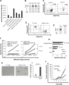
Cervical cancer is a major cause of cancer death in females worldwide. Cervical cancer stem-like cells (CSCs)/cancer-initiating cells (CICs) are resistant to conventional radiotherapy and chemotherapy, and CSCs/CICs are thought to be responsible for recurrence. Eradication of CSCs/CICs is thus essential to cure cervical cancer. In this study, we isolated cervical CSCs/CICs by sphere culture, and we identified a cancer testis (CT) antigen, CTCFL/BORIS, that is expressed in cervical CSCs/CICs. BORIS has 23 mRNA isoform variants classified by 6 subfamilies (sfs), and they encode 17 different BORIS peptides. BORIS sf1 and sf4 are expressed in both CSCs/CICs and non-CSCs/CICs, whereas BORIS sf6 is expressed only in CSCs/CICs. Overexpression of BORIS sf6 in cervical cancer cells increased sphere formation and tumor-initiating ability compared with those in control cells, whereas overexpression of BORIS sf1 and BORIS sf4 resulted in only slight increases. Thus, BORIS sf6 is a cervical CSC/CIC-specific subfamily and has a role in the maintenance of cervical CSCs/CICs. BORIS sf6 contains a specific c-terminal domain (C34), and we identified a human leukocyte antigen (HLA)-A2-restricted antigenic peptide, BORIS C34_24(9) encoded by BORIS sf6. A BORIS C34_24(9)-specific cytotoxic T cell (CTL) clone showed cytotoxicity for BORIS sf6-overexpressing cervical cancer cells. Furthermore, the CTL clone significantly suppressed sphere formation of CaSki cells. Taken together, the results indicate that the CT antigen BORIS sf6 is specifically expressed in cervical CSCs/CICs, that BORIS sf6 has a role in the maintenance of CSCs/CICs, and that BORIS C34_24(9) peptide is a promising candidate for cervical CSC/CIC-targeting immunotherapy.
Keywords: BORIS; CTL; cancer stem cells; cervical cancer; peptide vaccine.
Conflict of interest statement
The authors have no financial conflicts of interest.
Figures


Similar articles
-
Brother of the regulator of the imprinted site (BORIS) variant subfamily 6 is a novel target of lung cancer stem-like cell immunotherapy.PLoS One. 2017 Mar 1;12(3):e0171460. doi: 10.1371/journal.pone.0171460. eCollection 2017. PLoS One. 2017. PMID: 28248963 Free PMC article.
-
Identification and functional analysis of variants of a cancer/testis antigen LEMD1 in colorectal cancer stem-like cells.Biochem Biophys Res Commun. 2017 Apr 8;485(3):651-657. doi: 10.1016/j.bbrc.2017.02.081. Epub 2017 Feb 20. Biochem Biophys Res Commun. 2017. PMID: 28219643
-
Annexin A3 as a potential target for immunotherapy of liver cancer stem-like cells.Stem Cells. 2015 Feb;33(2):354-66. doi: 10.1002/stem.1850. Stem Cells. 2015. PMID: 25267273
-
Targeting CTCFL/BORIS for the immunotherapy of cancer.Cancer Immunol Immunother. 2018 Dec;67(12):1955-1965. doi: 10.1007/s00262-018-2251-8. Epub 2018 Nov 2. Cancer Immunol Immunother. 2018. PMID: 30390146 Free PMC article. Review.
-
Cancer stem cells (CSCs), cervical CSCs and targeted therapies.Oncotarget. 2017 May 23;8(21):35351-35367. doi: 10.18632/oncotarget.10169. Oncotarget. 2017. PMID: 27343550 Free PMC article. Review.
Cited by
-
Establishment and evaluation of cell and animal models expressing BORIS subfamily 2 variant.Ann Transl Med. 2022 Jun;10(11):632. doi: 10.21037/atm-21-6336. Ann Transl Med. 2022. PMID: 35813346 Free PMC article.
-
Brother of the regulator of the imprinted site (BORIS) variant subfamily 6 is a novel target of lung cancer stem-like cell immunotherapy.PLoS One. 2017 Mar 1;12(3):e0171460. doi: 10.1371/journal.pone.0171460. eCollection 2017. PLoS One. 2017. PMID: 28248963 Free PMC article.
-
Tumor-infiltrating CD8+ T cells recognize a heterogeneously expressed functional neoantigen in clear cell renal cell carcinoma.Cancer Immunol Immunother. 2022 Apr;71(4):905-918. doi: 10.1007/s00262-021-03048-6. Epub 2021 Sep 7. Cancer Immunol Immunother. 2022. PMID: 34491407 Free PMC article.
-
BTApep-TAT peptide inhibits ADP-ribosylation of BORIS to induce DNA damage in cancer.Mol Cancer. 2022 Aug 2;21(1):158. doi: 10.1186/s12943-022-01621-w. Mol Cancer. 2022. PMID: 35918747 Free PMC article.
-
Immunoprotective effect of an in silico designed multiepitope cancer vaccine with BORIS cancer-testis antigen target in a murine mammary carcinoma model.Sci Rep. 2021 Nov 30;11(1):23121. doi: 10.1038/s41598-021-01770-w. Sci Rep. 2021. PMID: 34848739 Free PMC article.
References
-
- Jemal A, Bray F, Center MM, Ferlay J, Ward E, Forman D. Global cancer statistics. CA Cancer J Clin. 2011;61:69–90. - PubMed
-
- Baldwin P, Laskey R, Coleman N. Translational approaches to improving cervical screening. Nat Rev Cancer. 2003;3:217–226. - PubMed
-
- Koh WJ, Greer BE, Abu-Rustum NR, Apte SM, Campos SM, Chan J, Cho KR, Cohn D, Crispens MA, DuPont N, Eifel PJ, Gaffney DK, Giuntoli RL, et al. Cervical cancer. Journal of the National Comprehensive Cancer Network. 2013;11:320–343. - PubMed
-
- Visvader JE, Lindeman GJ. Cancer stem cells in solid tumours: accumulating evidence and unresolved questions. Nat Rev Cancer. 2008;8:755–768. - PubMed
-
- Hirohashi Y, Torigoe T, Inoda S, Takahashi A, Morita R, Nishizawa S, Tamura Y, Suzuki H, Toyota M, Sato N. Immune response against tumor antigens expressed on human cancer stem-like cells/tumor-initiating cells. Immunotherapy. 2010;2:201–211. - PubMed
MeSH terms
Substances
LinkOut - more resources
Full Text Sources
Other Literature Sources
Medical
Research Materials
Miscellaneous

