. 2016 May;24(5):877-89.
doi: 10.1038/mt.2016.32. Epub 2016 Feb 5.

Gene Transfer of Brain-derived Neurotrophic Factor (BDNF) Prevents Neurodegeneration Triggered by FXN Deficiency

Affiliations

Gene Transfer of Brain-derived Neurotrophic Factor (BDNF) Prevents Neurodegeneration Triggered by FXN Deficiency

Yurika Katsu-Jiménez et al. Mol Ther. 2016 May.

Abstract

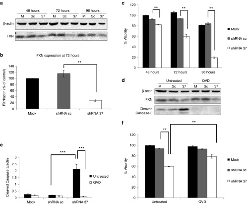
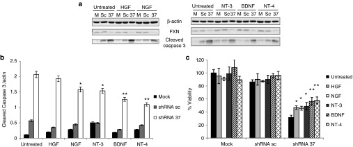
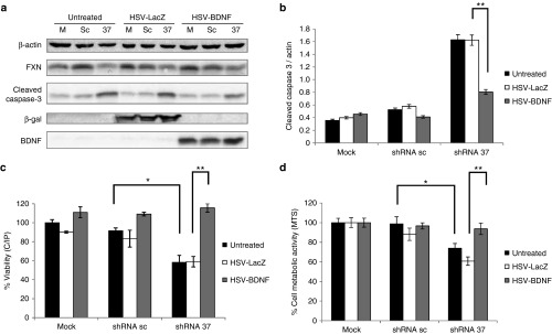
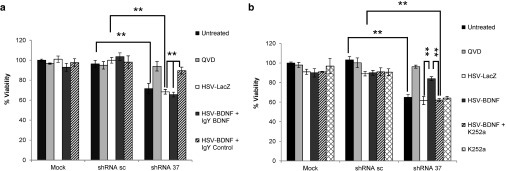
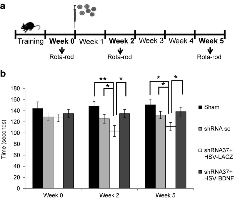
Friedreich's ataxia is a predominantly neurodegenerative disease caused by recessive mutations that produce a deficiency of frataxin (FXN). Here, we have used a herpesviral amplicon vector carrying a gene encoding for brain-derived neurotrophic factor (BDNF) to drive its overexpression in neuronal cells and test for its effect on FXN-deficient neurons both in culture and in the mouse cerebellum in vivo. Gene transfer of BDNF to primary cultures of mouse neurons prevents the apoptosis which is triggered by the knockdown of FXN gene expression. This neuroprotective effect of BDNF is also observed in vivo in a viral vector-based knockdown mouse cerebellar model. The injection of a lentiviral vector carrying a minigene encoding for a FXN-specific short hairpin ribonucleic acid (shRNA) into the mouse cerebellar cortex triggers a FXN deficit which is accompanied by significant apoptosis of granule neurons as well as loss of calbindin in Purkinje cells. These pathological changes are accompanied by a loss of motor coordination of mice as assayed by the rota-rod test. Coinjection of a herpesviral vector encoding for BDNF efficiently prevents both the development of cerebellar neuropathology and the ataxic phenotype. These data demonstrate the potential therapeutic usefulness of neurotrophins like BDNF to protect FXN-deficient neurons from degeneration.


Figures

Figure 1
Figure 1
Knockdown of FXN in cultured neurons triggers apoptotic cell death. Primary cortical neurons were transduced with lentivectors encoding for shRNA sequences against human FXN (shRNA-37), or containing a random scrambled shRNA sequence (shRNA-sc), or were left untransduced as an untreated control. (a) Representative western-blot analysis of FXN expression at 48, 72, and 96 hours post-transduction in comparison with that found in untransduced cells (mock, m). (b) Densitometric analysis of FXN levels in cultured neurons reveals significant differences at 72 hours post-transduction. (c) Cell viability of cultured neurons was estimated at 48, 72, and 96 hours post-transduction. (d) Western-blot analysis of FXN and cleaved caspase-3 at 72 hours post-transduction in the absence or presence of 50 µM Q-VD-OPh (N-(2-Quinolyl)-valyl-aspartyl-(2,6-difluorophenoxy)methyl ketone). (e) Quantification of cleaved caspase-3 levels in FXN-deficient neurons shows significant differences at 72 hours post-transduction. (f) Cell viability of cultured neurons at 72 hours post-transduction in the absence or presence of 50 µM Q-VD-OPh (QVD). Significant differences were shown between FXN-deficient neurons with or without QVD. Data represent mean values ± SEM from three independent experiments, **P ≤ 0.005, ***P ≤ 0.0005.
Figure 2
Figure 2
Effects of recombinant human neurotrophic factors in FXN-deficient neurons in culture. Primary cortical neurons were transduced with lentivectors (shRNA-sc or shRNA-37) or were left untransduced as a mock control (m), in the presence of different trophic factors such as hepatocyte growth factor (HGF), neurotrophin-3 (NT-3), neurotrophin-4 (NT-4) and brain-derived neurotrophic factor (BDNF) (100 ng/ml) and nerve growth factor (NGF) (1 µg/ml). (a) Representative western-blot analysis of FXN and cleaved caspase-3 levels at 72 hours upon transduction and treatments. (b) Quantification of cleaved caspase-3 levels in FXN-deficient neurons upon transduction reveals a decrease of caspase-3 activation when cells were treated with neurotrophins compared to untreated cells. (c) Cell viability of cultured neurons at 72 hours upon transduction shows significant differences between FXN-deficient neurons with neurotrophins compared to untreated cells. Data represent mean values ± SEM from three independent experiments, *P ≤ 0.05, **P ≤ 0.005.
Figure 3
Figure 3
BDNF gene transfer prevents apoptotic cell death of FXN-deficient neurons in culture. Primary cortical neurons were transduced with lentivectors (shRNA-sc or shRNA-37) or were left untransduced as a mock control (m), in the presence of HSV-1 amplicon vectors containing the cDNA of BDNF (HSV-BDNF) or the cDNA of β-galactosidase (HSV-LacZ) as control. (a) Representative western-blot analysis of FXN, cleaved caspase-3, β-galactosidase, and BDNF levels at 72 hours upon cotransduction. (b) Quantification of cleaved caspase-3 levels in FXN-deficient neurons upon cotransduction reveals a decrease of caspase-3 activation when BDNF was overexpressed. (c) Cell viability of cultured neurons at 72 hours upon cotransduction as assessed by the calcein/propidium iodide assay shows significant differences between FXN-deficient neurons with or without HSV-BDNF. (d) Cell metabolic activity of cultured neurons at 72 hours upon cotransduction was assessed by MTS reduction and also shows significant differences between FXN-deficient neurons with or without HSV-BDNF. Data represent mean values ± SEM from three independent experiments, *P ≤ 0.05, **P ≤ 0.005.
Figure 4
Figure 4
BDNF neuroprotective effect is mediated by TrkB receptor. Primary cortical neurons were cotransduced with lentivectors (shRNA-sc or shRNA-37) and HSV-1 amplicon vectors (HSV-LacZ or HSV-BDNF). (a) Cell viability assay of cultured neurons at 72 hours upon cotransduction shows significant difference in the presence of the BDNF neutralizing antibody or a control IgY antibody (20 µg/ml) added to the media. (b) Cell viability assay of cultured neurons at 72 hours upon cotransduction shows significant difference in the absence or presence of 50 nM of TrkB inhibitor K252a. Data represent mean values ± SEM from three independent experiments, **P ≤ 0.005.
Figure 5
Figure 5
FXN knockdown and BDNF overexpression after stereotaxic injection of viral vectors into the cerebellum. 8-week-old male C57BL/6 mice were injected with lentivectors (shRNA-sc or shRNA-37) and HSV-1 amplicon vectors (HSV-LacZ or HSV-BDNF) by stereotaxic injection into the cerebellum. (a) Schematic sagittal section of mouse cerebellum at coordinates AP −6.5 mm; lateral 1 mm; V −2.5 mm. (b) Coronal cerebellar section staining with nucleus marker 4́-6-diamidine-2-phenylindole (DAPI) (white) that indicates the region injected. Bar: 10 µm. (c) Confocal images show representative staining of cells, after immunohistochemistry for β-galactosidase (red) and the nucleus marker DAPI (blue), in the cerebellar cortex of sham animals or treated with shRNA-sc or with shRNA-37 and HSV-LacZ or with shRNA-37 and HSV-BDNF. Cerebellar sections were from mice killed 4 days after injection. Bar: 20 µm. (d) Scheme shows the four experimental groups of animals, sham, sc, 37 + HSV-LacZ, and 37 + HSV-BDNF. (e) Western-blot analysis of FXN levels in cerebella of mice 4 days after injection. (f) Densitometric analysis of the levels of FXN in cerebellum reveals a significant decreased in those animals injected with shRNA-37. (g) BDNF levels in cerebella of mice 4 days after injection were measured by enzyme linked immunosorbent assay ELISA. Data represent mean values ± SEM from three independent experiments, *P ≤ 0.05, **P ≤ 0.005.
Figure 6
Figure 6
BDNF prevents apoptotic cell death of FXN-deficient cerebellar neurons in vivo. Animals were co-transduced with concentrated lentivectors (shRNA-sc or shRNA-37) and HSV-1 amplicon vectors (HSV-LacZ or HSV-BDNF) by in vivo stereotaxic injection into the cerebellum. (a) Confocal images show representative staining of cells after immunohistochemistry for fractin (red) and the nucleus marker DAPI (blue), in the cerebellum of sham animals or treated with shRNA-sc or with shRNA-37 and HSV-LacZ or with shRNA-37 and HSV-BDNF. Cerebellar sections were from mice killed 4 days after injection. Yellow arrows indicate positive cell for fractin. Bar: 10 µm. (b) Quantification of fractin-positive cells in FXN-deficient cerebella indicated a significant decrease when BDNF was overexpressed compared with animals treated with HSV-LacZ. (c) Representative western-blot of PARP 1 and cleaved caspase-3 levels in cerebella of mice killed 4 days after injection. (d) Quantification of cleaved caspase-3 levels in FXN-deficient cerebella indicated a significant decrease when BDNF was overexpressed in comparison with animals treated with HSV-LacZ. (e) Quantification of cleaved PARP 1 levels in FXN-deficient cerebella indicated a significant decrease when BDNF was overexpressed compared with animals treated with HSV-LacZ. Data represent mean values ± SEM from three independent experiments, *P ≤ 0. 05, **P ≤ 0.005.
Figure 7
Figure 7
BDNF prevents the loss of calbindin in FXN-deficient cerebellar Purkinje cell in vivo. Animals were cotransduced with lentivectors (shRNA-sc or shRNA-37), and HSV-1 amplicon vectors (HSV-LacZ or HSV-BDNF) by in vivo stereotaxic injection into the cerebellum. (a) Confocal images show representative staining of cells, after immunohistochemistry for calbindin 28k (red) and the nucleus marker DAPI (blue), in the cerebellum of sham animals or treated with shRNA-sc or with shRNA-37 and HSV-LacZ or with shRNA-37 and HSV-BDNF. Cerebellar sections were from mice killed 4 days after injection. Bar: 20 µm. (b) Representative western-blot of calbindin 28k levels in cerebella of mice killed 4 days after injection. (c) Quantification of calbindin 28k levels in FXN-deficient cerebella indicated a significant increase when BDNF was overexpressed in comparison with animals treated with HSV-LacZ. Data represent mean values ± SEM from three independent experiments, **P ≤ 0.005.
Figure 8
Figure 8
Rota-rod performance in FXN knockdown mice by in vivo stereotaxic injection of viral vectors into the cerebellum. Animals were co-transduced with lentivectors (shRNA-sc or shRNA-37) and HSV-1 amplicon vectors (HSV-LacZ or HSV-BDNF) by in vivo stereotaxic injection into the cerebellum. (a) Scheme of experimental procedure. (b) Mice were analyzed in the accelerating rota-rod test to measure motor activity and coordination, first before injection (week 0) and at 2 and 5 weeks after stereotaxic injection in the cerebellum. Data represent mean values ± SEM from 10 mice, *P ≤ 0. 05, **P ≤ 0.005.

Comment in

Similar articles

Cited by

References

    1. Parkinson, MH, Boesch, S, Nachbauer, W, Mariotti, C and Giunti, P (2013). Clinical features of Friedreich's ataxia: classical and atypical phenotypes. J Neurochem 126(Suppl 1): 103–117. - PubMed
    1. Delatycki, MB and Corben, LA (2012). Clinical features of Friedreich ataxia. J Child Neurol 27: 1133–1137. - PMC - PubMed
    1. Lynch, DR, Deutsch, EC, Wilson, RB and Tennekoon, G (2012). Unanswered questions in Friedreich ataxia. J Child Neurol 27: 1223–1229. - PMC - PubMed
    1. Marmolino, D (2011). Friedreich's ataxia: past, present and future. Brain Res Rev 67: 311–330. - PubMed
    1. Koeppen, AH and Mazurkiewicz, JE (2013). Friedreich ataxia: neuropathology revised. J Neuropathol Exp Neurol 72: 78–90. - PMC - PubMed

Publication types

Substances