. 2016 Feb 5;11(2):e0148633.
doi: 10.1371/journal.pone.0148633. eCollection 2016.

Voltage-Gated K+ Channel, Kv3.3 Is Involved in Hemin-Induced K562 Differentiation

Affiliations

Voltage-Gated K+ Channel, Kv3.3 Is Involved in Hemin-Induced K562 Differentiation

Min Seok Song et al. PLoS One. .

Abstract

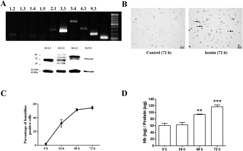
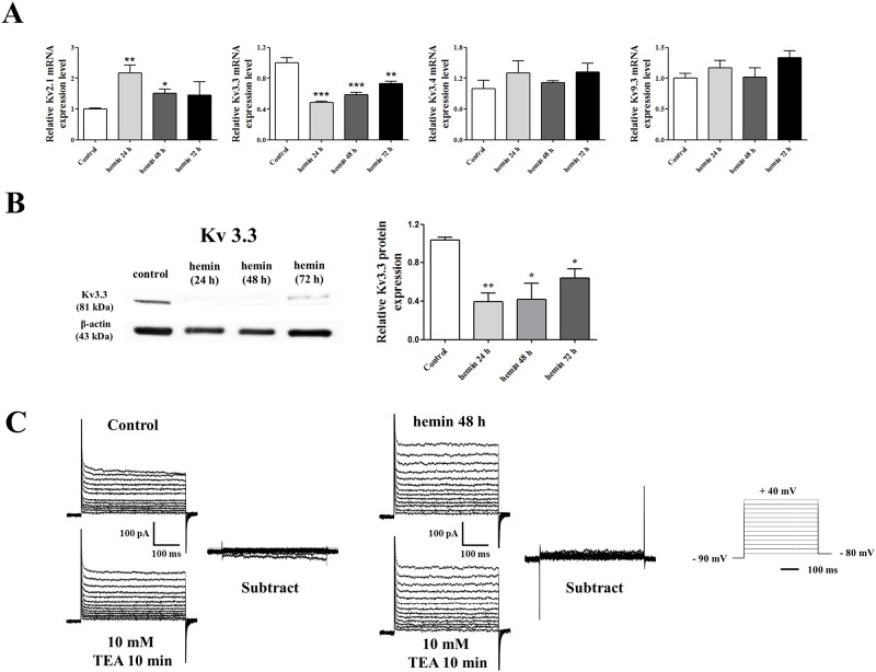
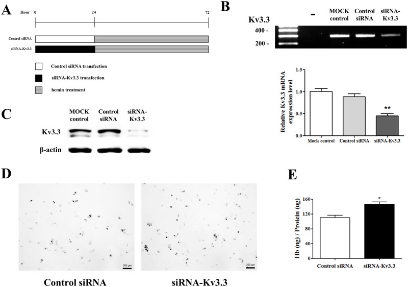
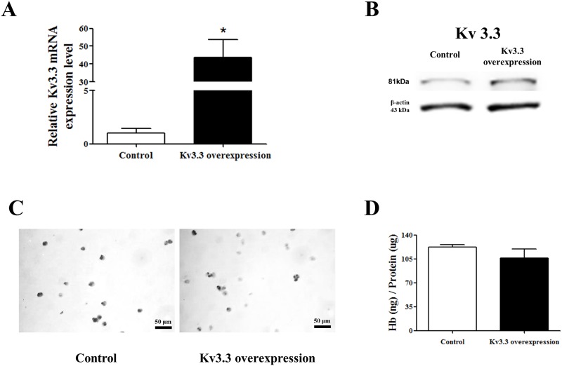
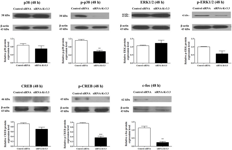
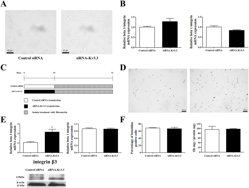
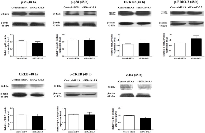
Voltage-gated K+ (Kv) channels are well known to be involved in cell proliferation. However, even though cell proliferation is closely related to cell differentiation, the relationship between Kv channels and cell differentiation remains poorly investigated. This study demonstrates that Kv3.3 is involved in K562 cell erythroid differentiation. Down-regulation of Kv3.3 using siRNA-Kv3.3 increased hemin-induced K562 erythroid differentiation through decreased activation of signal molecules such as p38, cAMP response element-binding protein, and c-fos. Down-regulation of Kv3.3 also enhanced cell adhesion by increasing integrin β3 and this effect was amplified when the cells were cultured with fibronectin. The Kv channels, or at least Kv3.3, appear to be associated with cell differentiation; therefore, understanding the mechanisms of Kv channel regulation of cell differentiation would provide important information regarding vital cellular processes.


Conflict of interest statement

Competing Interests: The authors have declared that no competing interests exist.

Figures

Fig 1
Fig 1. Identification of Kv channels in K562 cells and the erythroid differentiation of K562 cells using hemin.
(A) RT-PCR data analysis demonstrated 7 different subtypes of Kv channels (Kv1.2, Kv1.4, Kv1.5, Kv2.1, Kv3.3, Kv3.4, Kv4.3, and Kv9.3). Western blot demonstrated the protein expression of Kv1.3, Kv2.1, Kv3.3, and Kv9.3. (B) K562 cells differentiated into erythroid cells were stained with benzidine after 72 h of differentiation using hemin (magnification ×40). Benzidine-positive cells appeared black, indicated by colored arrows. (C) The percentage of benzidine-positive cells was counted at 4 different time points (0, 24, 48, and 72 h). (D) The hemoglobin content of the differentiated K562 cells was measured at each indicated time point using a modified QuantiChrom Heme Assay. The concentration of hemoglobin at each time point was expressed as nanograms of heme per microgram of total protein.
Fig 2
Fig 2. The expression of Kv channels and whole cell patch clamp recording during the late stage of differentiation.
(A) After hemin-induced K562 cell differentiation, the mRNA expression levels of Kv channels, including Kv2.1, Kv3.3, Kv3.4, and Kv9.3, were compared with those of undifferentiated cells. The mRNA expression level of Kv2.1 was increased, whereas the mRNA expression level of Kv3.3 was decreased during erythroid differentiation. The mRNA expression of Kv3.4 and Kv9.3 were not altered during erythroid differentiation. The relative mRNA expressions of the Kv channels were normalized to the GAPDH gene and expressed as a fold change relative to the Mock control group. (B) The protein level of Kv3.3 decreased significantly after 24 hours of hemin-induced erythroid differentiation in K562 cells. The relative protein expression of the Kv3.3 was expressed as a fold change relative to the control group. (C) Representative current traces recorded from K562 cells. There was no TEA-sensitive current before and after hemin-induced erythroid differentiation. Experiments were performed in triplicate, and data are expressed as mean ± standard error. **p<0.01 compared with control value.
Fig 3
Fig 3. Silenced Kv3.3 increased hemin-induced K562 cell differentiation.
Hemin-induced K562 cell differentiation was significantly increased by decreasing the Kv3.3 expression using siRNA-Kv3.3. (A) Protocol for control siRNA and siRNA-Kv3.3 transfection and hemin treatment. After 24 h of transfection, the K562 cells were cultured with hemin for 48 h. mRNA (B) and protein (C) expressions were suppressed when the cells were transfected with siRNA-Kv3.3. RT-PCR was performed after 48 h of transfection. The relative mRNA expression of the Kv3.3 was normalized to the GAPDH gene and expressed as a fold change relative to the Mock control group. (D) Benzidine staining demonstrated greater hemoglobin formation in siRNA-Kv3.3-transfected cells (right) compared to control siRNA-transfected cells (left) during hemin-induced K562 cell erythroid differentiation (magnification ×40). Staining was performed after 24 h of transfection and 24 h of differentiation. (E) The amounts of hemoglobin content were increased by siRNA-Kv3.3 transfection compared to control. The hemoglobin content was increased by about 50% by siRNA-Kv3.3 transfection. Experiments were performed in triplicate, and data are expressed as mean ± standard error. *p<0.05 compared with control value.
Fig 4
Fig 4. Overexpression of Kv3.3 did not have a clear effect on hemin-induced erythroid differentiation.
mRNA (A) and protein (B) expression levels of Kv3.3 were increased in the K562 overexpressed cell line compared to the normal K562 cell line. The relative mRNA expression of the Kv3.3 was normalized to the GAPDH gene and expressed as a fold change relative to the control group. The relative protein expressions of the Kv3.3 were expressed as a fold change relative to the control. (C) The degree of hemin-induced erythroid differentiation was measured using benzidine staining in Kv3.3-overexpressed K562 cells or normal K562 cells. There was no significant difference between the two, however. (D) Hemoglobin quantification demonstrated that overexpressed Kv3.3 did not have any effect on hemin-induced K562 erythroid differentiation.
Fig 5
Fig 5. Signaling mechanisms of K562 erythroid differentiation regulation by siRNA- Kv3.3 transfection.
The expressions of p38, ERK1/2, CREB, and c-fos during cell differentiation changed with siRNA-Kv3.3 transfection. The levels of the phosphorylated, activated forms of p38 and CREB were lower in siRNA-Kv3.3 transfected cells than in control cells. The levels of c-fos during cell differentiation were also reduced after siRNA- Kv3.3 transfection. The levels of phosphorylated ERK2 (p-42 MAPK) also seemed lower; however, the differences were not statistically significant. Phosphorylated ERK1 (p-44 MAPK) was not detected. No changes were noted for the total p38, total ERK1/2, and total CREB. The graphs show the quantitative analysis of each protein. Western blot assay was performed when transfected cells were differentiated for 48 h with hemin; each assay was performed in triplicate, and data are expressed as mean ± standard error. **p<0.01 compared with control value. The relative protein expressions of the signal molecules were expressed as a fold change relative to the control group.
Fig 6
Fig 6. The relationship between Kv3.3 and the early stage of K562 erythroid differentiation.
(A) The mRNA expression levels of the Kv channels, including Kv2.1, Kv3.3, Kv3.4, and Kv9.3, did not correlate with the hemin-induced K562 erythroid differentiation at the indicated time points (10 min, 30 min, and 1 h). The relative mRNA expressions of the Kv channels were normalized to the GAPDH gene and expressed as a fold change relative to the control group. (B) The protein expression level of Kv3.3 was estimated after inducing 10 min and 30 min of erythroid differentiation, and there was no change compared to the control cells. The relative protein expressions of the Kv3.3 were expressed as a fold change relative to the control.
Fig 7
Fig 7. In the early stage of K562 erythroid differentiation, Kv3.3 knockdown using siRNA-Kv3.3 did not have any effect on the expression levels of signaling molecules involved in K562 erythroid differentiation.
The protein expression levels of p38 and ERK were measured after 24 h of siRNA-Kv3.3 transfection and 10 min or 30 min of erythroid differentiation. During the early stage of erythroid differentiation, down-regulated Kv3.3 had no effect on the protein levels of p38 and ERK. The relative protein expressions of the signal molecules were expressed as a fold change relative to the control siRNA group.
Fig 8
Fig 8. Effects of siRNA-Kv3.3 transfection on cell adhesion.
(A) Attached K562 cells were detected in siRNA-Kv3.3-transfected cell cultures (right), whereas fewer attached cells were found in control cultures (left) (magnification ×200). (B) The mRNA expression levels of integrin β3 (left) and integrin β1 (right) during hemin-induced K562 cell erythroid differentiation by transfection of siRNA-Kv3.3. (C) The protocol for control siRNA and siRNA-Kv3.3 transfection and hemin treatment with fibronectin. After 24 h of transfection, the K562 cells were cultured with hemin in fibronectin-coated wells for 48 h. (D) Culturing the cells in fibronectin-coated wells (10 μg/ml) significantly improved cell adhesion during the hemin-induced erythroid differentiation of siRNA-Kv3.3-transfected cells (magnification ×40). (E) Cells cultured in fibronectin-coated wells showed amplified effects of decreased Kv3.3 on integrin β3 levels. The mRNA and protein expression levels of integrin β3 in siRNA-Kv3.3-transfected cells increased much more during hemin-induced K562 cell erythroid differentiation than in the control group (*p<0.05). On the other hand, no differences were noted in integrin β1 expression between the control and siRNA-Kv3.3 transfected-cells when the cells were cultured in fibronectin-coated wells. (F) Benzidine staining (left) and hemoglobin quantification (right) indicated that increased erythroid differentiation by siRNA-Kv3.3 was not detected when the cells were cultured in fibronectin-coated wells. The relative mRNA expressions of the integrins were normalized to the GAPDH gene and expressed as a fold change relative to the control siRNA group.
Fig 9
Fig 9. Signaling mechanisms of K562 differentiation regulation by siRNA-Kv3.3 transfection and cultures in fibronectin plates.
No changes in the expression levels of p38, ERK1/2, CREB, and c-fos were noted in response to decreased Kv3.3 levels compared to control cells during cell differentiation in fibronectin-coated wells. No differences were noted in the total amounts or amounts of phosphorylated forms of ERK1/2, p38, and CREB expression in the control and siRNA-transfected cells. No differences were observed in the c-fos levels following siRNA-Kv3.3 transfection when the cells were cultured with fibronectin. The graphs indicate the quantitative analysis of each protein. Western blot assays were performed when the transfected cells were differentiated for 48 h with hemin and fibronectin. Each assay was performed in triplicate, and data are expressed as mean ± standard error. The relative protein expressions of the signal molecules were expressed as a fold change relative to the control siRNA group.

Similar articles

Cited by

References

    1. Hille B. Ion channels of excitable membranes. 3rd ed Sunderland: Sinauer associates, Inc; 2001.
    1. Pardo LA, Suhmer W. Eag1 as a cancer target. Expert opinion on therapeutic targets. 2008;12(7):837–43. Epub 2008/06/17. 10.1517/14728222.12.7.837 . - DOI - PubMed
    1. Bardou O, Trinh NT, Brochiero E. Molecular diversity and function of K+ channels in airway and alveolar epithelial cells. American journal of physiology Lung cellular and molecular physiology. 2009;296(2):L145–55. Epub 2008/12/09. 10.1152/ajplung.90525.2008 . - DOI - PubMed
    1. Kunzelmann K. Ion channels and cancer. J Membrane Biol. 2005;205(3):159–73. 10.1007/s00232-005-0781-4 ISI:000233908900007. - DOI - PubMed
    1. Lang F, Foller M, Lang KS, Lang PA, Ritter M, Gulbins E, et al. Ion channels in cell proliferation and apoptotic cell death. J Membrane Biol. 2005;205(3):147–57. 10.1007/s00232-005-0780-5 ISI:000233908900006. - DOI - PubMed

Publication types

MeSH terms