Review
. 2016 May;137(3):312-30.
doi: 10.1111/jnc.13564. Epub 2016 Mar 3.

Dynamic DNA methylation controls glutamate receptor trafficking and synaptic scaling

Affiliations
Review

Dynamic DNA methylation controls glutamate receptor trafficking and synaptic scaling

J David Sweatt. J Neurochem. 2016 May.

Abstract

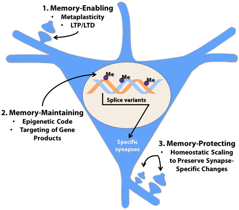
Hebbian plasticity, including long-term potentiation and long-term depression, has long been regarded as important for local circuit refinement in the context of memory formation and stabilization. However, circuit development and stabilization additionally relies on non-Hebbian, homeostatic, forms of plasticity such as synaptic scaling. Synaptic scaling is induced by chronic increases or decreases in neuronal activity. Synaptic scaling is associated with cell-wide adjustments in postsynaptic receptor density, and can occur in a multiplicative manner resulting in preservation of relative synaptic strengths across the entire neuron's population of synapses. Both active DNA methylation and demethylation have been validated as crucial regulators of gene transcription during learning, and synaptic scaling is known to be transcriptionally dependent. However, it has been unclear whether homeostatic forms of plasticity such as synaptic scaling are regulated via epigenetic mechanisms. This review describes exciting recent work that has demonstrated a role for active changes in neuronal DNA methylation and demethylation as a controller of synaptic scaling and glutamate receptor trafficking. These findings bring together three major categories of memory-associated mechanisms that were previously largely considered separately: DNA methylation, homeostatic plasticity, and glutamate receptor trafficking. This review describes exciting recent work that has demonstrated a role for active changes in neuronal DNA methylation and demethylation as a controller of synaptic scaling and glutamate receptor trafficking. These findings bring together three major categories of memory-associated mechanisms that were previously considered separately: glutamate receptor trafficking, DNA methylation, and homeostatic plasticity.

Keywords: AMPA receptor; TET; active demethylation; epigenetic; homeostatic plasticity; memory.


Figures

Figure 1
Figure 1. Biochemical pathway for modification of cytosine within DNA by TET
(A) 5-methylCytosine (5mC) bases, introduced by DNA methyltransferase (DNMT) enzymes, can subsequently be oxidized iteratively by the TET family of dioxygenases to 5-hydroxymethylCytosine (5hmC), 5-formylCytosine (5fC) and 5-carboxylated Cytosine (5caC). Figure and legend adapted from R.M. Kohli and Y Zhang, ‘TET enzymes, TDG and the dynamics of DNA demethylation’ Nature 502, 472–479 (24 October 2013). (B) TET1 dioxygenase coverts 5-methylcytosine (5mC) to 5-hydroxymethylcytosine (5hmC) in mammalian cells. Subsequent demethylation occurs through a process that requires deamination and the base-excision pathway (BER) mechanism. Demethylation of both exogenously introduced and endogenous 5hmCs is promoted by the AID (activation-induced deaminase)/APOBEC (apolipoprotein B mRNA-editing enzyme complex) family of cytidine deaminases. Furthermore, Tet1 and Apobec1 are involved in neuronal activity-induced, region-specific, active DNA demethylation and subsequent gene expression in the adult CNS. Figure and legend adapted from (Kohli & Zhang 2013, Guo et al. 2011c).
Figure 2
Figure 2. DNA methylation regulates synaptic scaling
(A) Homeostatic upscaling of excitatory synaptic strength due to chronic inhibition of sodium channels with tetrodotoxin (TTX). (i) Sample spontaneous miniature excitatory post-synaptic current traces (mEPSCs) – a sample recording epoch illustrating mEPSC records from cortical pyramidal neurons after 24 hours of exposure to vehicle control (CTL) or TTX (blue) is shown. Downward spikes are postsynaptic mEPSCs triggered by glutamate release presynaptically. (ii) Cumulative probability distributions of mEPSC amplitude and overall mean mEPSC amplitudes in control and TTX-treated pyramidal neuron cultures are shown – the rightward shift of the blue line and increase in total average mEPSC amplitude are indicative of synaptic upscaling. P < 0.001, Kolmogorov-Smirnov (K-S) test. Inset: *P < 0.001, Mann-Whitney (M-W) test. (iii) Mean mEPSC frequencies do not increase in cortical pyramidal neurons treated with TTX, suggesting (as has been previously demonstrated) that the increase in (ii) is not due to an increase in total number of synaptic connections on the postsynaptic neuron. Data from cells pooled from at least six experiments for each condition (CTL, n = 21 cells; TTX, n = 15 cells). (B) Induction of excitatory synaptic scaling by TTX is blocked by DNMT inhibition. (i) Sample mEPSC traces from cortical pyramidal neurons after 24 hours of exposure to control (CTL) or TTX + the competitive DNMT inhibitor RG108 (green). (ii) Cumulative probability distributions and mean mEPSC amplitudes from cortical pyramidal neurons treated with TTX in the presence of RG108. Data are cumulative of cells pooled from at least three experiments for each condition (CTL, n = 17 cells; RG108 + TTX, n = 12 cells). These results compared to the data presented in (A) indicate that DNMT activity is necessary for TTX-induced synaptic scaling. (C) DNMT inhibition with RG108 or via DNMT mRNA knockdown upscales excitatory synaptic strength. (i) Sample mEPSC records from cortical pyramidal neurons after 24-hour treatment with control (CTL) or the DNMT inhibitor RG108 (red). (ii) Cumulative probability distributions and mean mEPSC amplitudes from cortical pyramidal neurons treated with RG108. These data indicate that DNA cytosine demethylation (known to occur secondary to DNMT inhibition) is capable of triggering synaptic upscaling. Please note that the data presented in (B) also indicate that the RG108-triggered synaptic upscaling is dependent on neuronal action potential firing, that is, that the RG108 effect is dependent on neuronal activity. Data from cells pooled from at least four experiments for each condition (CTL, n = 12 cells; RG108, n = 12 cells). P < 0.001, K-S test. Inset: *P < 0.001, M-W test. (D) Combined Dnmt1 and Dnmt3a knockdown multiplicatively upscales excitatory strength. Left Panel - Rank Order Plot of one thousand randomly selected mEPSC amplitudes from scrambled CTL and combined ASO treatment (orange values). Linear regression yielded a scaling factor of 1.34. Right Panel – Scaled-down amplitudes from combined ASO treatment were not different than scrambled CTL (K – S test, P = 0.1323). This graphical analysis illustrates that the synaptic upscaling induced by DNMT knockdown is multiplicative in nature. This finding is consistent with the hypothesis that the upscaling maintained relative individual synaptic strengths across the entire population of neuronal synapses, that is that the fractional contribution of each individual synapse was preserved and the entire neuronal synaptic population was regulated in a proportional, coordinated fashion. Bar graphs are means ± SEM. Figure and legend adapted from (Meadows et al. 2015).
Figure 3
Figure 3. Tet3 expression regulates glutamatergic synaptic transmission and synaptic scaling
(A) Synaptic activity-dependent expression of Tet3 regulates glutamatergic synaptic transmission. In these experiments the effects of TET1 knockdown (left panel), TET2 knockdown (middle panel) and TET3 knockdown (right panel) were evaluated, and overall glutamatergic synaptic activity was quantitated as a percent of control. In all cases the effects of TET isoform knockdown were evaluated ether in the presence of sodium channel blockade (TTX, blue graphs) or GABA receptor blockade (Bicuculline, orange graphs). Tet3-knockdown neurons exhibited elevated glutamatergic synaptic transmission when GABA receptors were blocked (right panel). (B) Neurons overexpressing TET3 catalytic domain exhibit decreased glutamatergic synaptic transmission, and TET3 catalytic domain overexpression blocks TTX-induced synaptic upscaling. The left and right bottom panels illustrate that TET3 catalytic domain overexpression (EYFP/Tet3 OE) down-scales glutamatergic synaptic transmission relative to enhanced yellow fluorescent protein (EYFP) controls (red versus green bars). The upper panel presents cumulative probability curves demonstrating that TTX by itself triggers upscaling (gray bars), while TET1 catalytic domain (Tet1-CD) triggers downscaling (blue bars) and blocks the TTX-induced upscaling (aqua bars). The orange bars illustrate that the effects of TET1 catalytic domain are dependent on the activity of the enzyme, because they are not mimicked by transfection of a mutant catalytically inactive catalytic domain (Tet1-mCD). Values represent mean ± s.e.m. (*P < 0.05, **P < 0.01). Figure and legend adapted from (Yu et al. 2015). Please see (Yu et al. 2015) for additional details.
Figure 4
Figure 4. Persisting changes in DNA methylation support long-term memory stabilization in the dorsomedial prefrontal cortex
(A) Both DNA methylation and gene transcription change in the dmPFC, in a time-dependent fashion, following training for contextual fear conditioning. (i-iii), location of promoter CpG islands analyzed in mDIP assay for Egr1/Zif268 (i), reelin (ii) and calcineurin (iii). (iv) Relative to naïve controls, all treatment groups showed demethylation of Egr1/zif268 at all time points (1h: F(3, 18) = 22.99, P ≤ 0.001; 1 day: F(3, 19) = 14.99, P ≤ 0.001; and 7 days post-training: F(3, 25) = 15.50, P ≤ 0.001). * post hoc comparisons, P ≤ 0.001. (v) Relative to all treatment groups, reelin's promoter is hypermethylated in trained animals at all time points (1h: F(3, 18) = 29.05, P ≤ 0.001; 1 day: F(3, 19) = 6.63, P ≤ 0.005; and 7 days post-training: F(3, 25) = 10.58, P ≤ 0.001). * post hoc comparisons, P ≤ 0.05. 7 days post-training, reelin's hypermethylation is significantly less than at 1 hour (F(2, 20) = 6.36, P ≤ 0.01; #). (vi) Relative to all treatment groups, calcineurin's promoter is hypermethylated in trained animals 24 hours and 7 days post-training (1h: F(3, 18) = 0.27, P ≥ 0.05; 1 day: F(3, 19) = 5.73, P ≤ 0.01; and 7 days post-training: F(3, 25) = 33.52, P ≤ 0.001). * post hoc comparisons, P ≤ 0.05. 1 and 7 days post-training, calcineurin's hypermethylation is significantly greater than at 1 hour (F(2, 20) = 13.96, P ≤ 0.01; #). (vii) Egr1/zif268 transcript is elevated in all treatment groups 1 hour after training (relative to naïve controls—context: t4 = 3.11, shock: t4 = 5.10, context + shock: t4 = 3.02; * P ≤ 0.05) and following a retrieval test 7 days after training (relative to naïve controls—context: t4 = 6.95, shock: t4 = 3.12, context + shock: t6 = 6.59; * P ≤ 0.05) (relative to 7 day no retrieval groups—F(5, 27) = 11.97, # post hoc comparisons P ≤ 0.005). (viii) Relative to all treatment groups, reelin transcript is significantly lower in trained animals 1 hour after training (relative to naïve controls—t4 = -9.53, * P ≤ 0.005; relative to context and shock controls—F(2, 12) = 6.14, # P ≤ 0.05). Following a retrieval test 7 days after training, trained animals' reelin transcript is significantly lower than naïve controls (t6 = -4.00, * P ≤ 0.01). (ix) Calcineurin transcript is equivalent across all groups. N's = 4-8 animals per group. Figure and legend adapted from (Miller et al. 2010). Please see (Miller et al. 2010) for additional details.
Figure 5
Figure 5. DNMT inhibition disrupts hippocampal place field stability in vivo. Top panels
illustrate data obtained using in vivo multi-electrode recording techniques to quantitate place cell firing patterns, and to evaluate consistency of firing of a single hippocampal (dorsal CA1/CA3) place cell. For illustrative purposes data from a single control animal (and a single place cell) are shown. Pseudo-color images illustrate firing rates (blue = low, red = high). In this experiment the animal was placed sequentially onto a round circular maze, with each session separated by 10 minutes. As shown, there is a strong correlation of place cell firing between session 1 and session 2, when the animal is simply re-placed into the same familiar environment. However, when the same animal is placed into a new different environment (session 3), the place cell shows a different place field firing pattern. The dissimilarities in place cell firing can be quantitated using a Pearson correlation coefficient. The bar graph in the Bottom Panel presents Pearson correlation coefficient data testing the effects of the DNMT inhibitor zebularine on place cell firing pattern consistency in this type of place cell experiment (n=5 Long-Evans rats, p<0.05). Zebularine infusion ICV leads to a significant decrease in Pearson correlation coefficients for animals when they are re-placed into the familiar environment. These data are consistent with the idea that DNMT inhibition leads to a destabilization of place cell firing patterns. For a more complete description of these results please see (Roth et al. 2015).
Figure 6
Figure 6. Potential mechanisms through which DNA methylation might control memory formation and stabilization
This diagram summarizes the three main categories of mechanisms whereby cytosine methylation might contribute to memories being established and maintained, termed: 1. Memory-Enabling, 2. Memory-Maintaining, and 3. Memory-Protecting. Please see the main text for further discussion.

Similar articles

Cited by

References

    1. Abbott LF, Nelson SB. Synaptic plasticity: taming the beast. Nat Neurosci. 2000;3(Suppl):1178–1183. - PubMed
    1. Alarcon JM, Malleret G, Touzani K, Vronskaya S, Ishii S, Kandel ER, Barco A. Chromatin acetylation, memory, and LTP are impaired in CBP+/- mice: a model for the cognitive deficit in Rubinstein-Taybi syndrome and its amelioration. Neuron. 2004;42:947–959. - PubMed
    1. Baker-Andresen D, Ratnu VS, Bredy TW. Dynamic DNA methylation: a prime candidate for genomic metaplasticity and behavioral adaptation. Trends Neurosci. 2013;36:3–13. - PubMed
    1. Barnes CA, Suster MS, Shen J, McNaughton BL. Multistability of cognitive maps in the hippocampus of old rats. Nature. 1997;388:272–275. - PubMed
    1. Barrett RM, Wood MA. Beyond transcription factors: the role of chromatin modifying enzymes in regulating transcription required for memory. Learn Mem. 2008;15:460–467. - PMC - PubMed

Publication types

Substances

LinkOut - more resources