. 2016 Mar;73(3):117-30.
doi: 10.1002/cm.21281.

Molecular evolution of troponin I and a role of its N-terminal extension in nematode locomotion

Affiliations

Molecular evolution of troponin I and a role of its N-terminal extension in nematode locomotion

Dawn E Barnes et al. Cytoskeleton (Hoboken). 2016 Mar.

Abstract

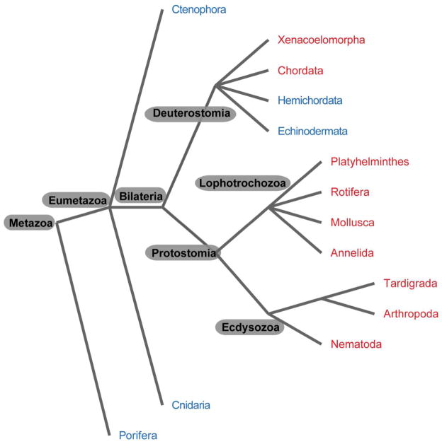
The troponin complex, composed of troponin T (TnT), troponin I (TnI), and troponin C (TnC), is the major calcium-dependent regulator of muscle contraction, which is present widely in both vertebrates and invertebrates. Little is known about evolutionary aspects of troponin in the animal kingdom. Using a combination of data mining and functional analysis of TnI, we report evidence that an N-terminal extension of TnI is present in most of bilaterian animals as a functionally important domain. Troponin components have been reported in species in most of representative bilaterian phyla. Comparison of TnI sequences shows that the core domains are conserved in all examined TnIs, and that N- and C-terminal extensions are variable among isoforms and species. In particular, N-terminal extensions are present in all protostome TnIs and chordate cardiac TnIs but lost in a subset of chordate TnIs including vertebrate skeletal-muscle isoforms. Transgenic rescue experiments in Caenorhabditis elegans striated muscle show that the N-terminal extension of TnI (UNC-27) is required for coordinated worm locomotion but not in sarcomere assembly and single muscle-contractility kinetics. These results suggest that N-terminal extensions of TnIs are retained from a TnI ancestor as a functional domain.

Keywords: actin; contraction; muscle; myosin; troponin.


Conflict of interest statement

The authors declare no conflict of interest.

Figures

Fig. 1
Fig. 1. Evolution of troponin
Phylogenetic relationships of 14 representative phyla in the animal kingdom are shown. Troponin-positive and –negative phyla are indicated by red and blue, respectively. Major taxonomic groups are indicated at or near branch points.
Fig. 2
Fig. 2. Molecular phylogenetic relationships of troponin I
Phylogenetic relationships of 19 TnI sequences from 9 species (8 phyla shown on the right) are shown. Three groups that match with major taxonomic groups are indicated by different colors: yellow (Protostomia, Ecdysozoa), pink (Protostomia, Lophotrochozoa), and green (Deuterostomia). N-terminal extensions are present in TnIs shown in red but absent in TnIs shown in blue. Entire sequence alignment is shown in Supplemental Fig. S1.
Fig. 3
Fig. 3. Comparison of structures of representative troponin I
(A) Schematic representation of structures of representative TnIs. N-terminal extension (NTE), four helices (H1 – H4), inhibitory region (IR), and C-terminal tail (extension) (C-tail) are shown. Major interaction sites with TnC, TnT, and actin are indicated on the top. (B) Sequence alignment of N-terminal extensions of five TnIs. Basic and acidic amino acids are shown in red and blue, respectively.
Fig. 4
Fig. 4. Effect of truncation of the N-terminal extension of troponin I on sarcomeric actin organization
(A–L), Micrographs of adult body wall muscle from wild-type (A–C), unc-27 (D–F), unc-27; GFP-UNC-27(WT) (G–I), or unc-27; GFP-UNC-27(ΔN) (J–L) are shown for filamentous (F-) actin stained with tetramethylrhodamine-phalloidin (left) and GFP (middle). Merged images are shown on the right column (F-actin in red and GFP in green). Bar, 50 μm. (M, N) Protein levels of TnI (M) and actin (N) were examined by Western blot. Lysates from 15 worms with indicated genotypes were loaded per sample and reacted with anti-Ascaris TnI antibody (M). Note that this antibody reacts with all four TnI isoforms, and showed significant reactivity with endogenous TnI with apparently higher molecular weight in the unc-27 mutant background as described previously (Obinata et al. 2010). (O, P) Quantitative analysis of the protein levels of GFP-UNC-27 (WT) and GFP-UNC-27(ΔN). Worm lysates (10 worms per sample) from unc-27; GFP-UNC-27(WT) and unc-27; GFP-UNC-27(ΔN) were subjected to Western blot with anti-GFP and anti-actin antibodies (O). Five samples for each strain and one sample for wild-type were examined. Band intensity of the GFP-fusion proteins was normalized by the actin levels and shown in the graph (P). Data are means ± standard errors, n = 5. Transgenically expressed GFP-UNC-27 (WT) and GFP-UNC-27(ΔN) were not at significantly different levels: n. s., not significant (P > 0.05).
Fig. 5
Fig. 5. Effect of truncation of the N-terminal extension of troponin I on worm motility in liquid
Worm motility of indicated strains was examined in liquid as beat frequency (beating per 30 sec). Data are means ± standard errors, n = 10. Results of one-way ANOVA are shown: n. s., not significant (P > 0.05); ***, P < 0.001.
Fig. 6
Fig. 6. Effect of truncation of the N-terminal extension of troponin I on bending amplitude during backward locomotion
Backward locomotion was induced by touching the head (A, C, E, and G). Then, amplitude of body bending (indicated as “A” in B) was measured and normalized by body length (indicated as “L” in B) (B, D, F, and H). Bar, 100 μm. Normalized bending amplitude (A/L) is shown in a box plot (I). Each box represents the 25th and 75th percentiles with a line at the median, and error bars indicate the 10th and 90th percentiles. n = 10. **, P < 0.001; ***, P < 0.0001.
Fig. 7
Fig. 7. Effect of truncation of the N-terminal extension of troponin I on rates of contraction and relaxation examined by optogenetics
(A) Representative images of an optogenetic experiment show that contraction of a worm expressing channelrhodopsin-2 in a microfluidic channel (60 μm wide) is induced by turning blue light on, and relaxation induced by turning blue light off. (B, C) Rate constants (s−1) for contraction (B) and relaxation (C) were measured from changes in the body area and expressed in box plots. n = 30. ***, P < 0.0001.

Similar articles

Cited by

References

    1. Arad M, Penas-Lado M, Monserrat L, Maron BJ, Sherrid M, Ho CY, Barr S, Karim A, Olson TM, Kamisago M, et al. Gene mutations in apical hypertrophic cardiomyopathy. Circulation. 2005;112:2805–2811. - PubMed
    1. Barbato JC, Huang QQ, Hossain MM, Bond M, Jin JP. Proteolytic N-terminal truncation of cardiac troponin I enhances ventricular diastolic function. J Biol Chem. 2005;280:6602–6609. - PubMed
    1. Biesiadecki BJ, Tachampa K, Yuan C, Jin JP, de Tombe PP, Solaro RJ. Removal of the cardiac troponin I N-terminal extension improves cardiac function in aged mice. J Biol Chem. 2010;285:19688–19698. - PMC - PubMed
    1. Blair JE, Hedges SB. Molecular phylogeny and divergence times of deuterostome animals. Mol Biol Evol. 2005;22:2275–2284. - PubMed
    1. Brenner S. The genetics of Caenorhabditis elegans. Genetics. 1974;77:71–94. - PMC - PubMed

Publication types

MeSH terms

LinkOut - more resources