. 1989 Dec 5;264(34):20422-9.

Human lamin B contains a farnesylated cysteine residue

Affiliations

Human lamin B contains a farnesylated cysteine residue

C C Farnsworth et al. J Biol Chem. .

Abstract

We recently showed that HeLa cell lamin B is modified by a mevalonic acid derivative. Here we identified the modified amino acid, determined its mode of linkage to the mevalonic acid derivative, and established the derivative's structure. A cysteine residue is modified because experiments with lamin B that had been biosynthetically labeled with [3H]mevalonic acid or [35S]cysteine and then extensively digested with proteases yielded 3H- or 35S-labeled products that co-chromatographed in five successive systems. A thioether linkage rather than a thioester linkage is involved because the mevalonic acid derivative could be released from the 3H-labeled products in a pentane-extractable form by treatment with Raney nickel but not with methanolic KOH. The derivative is a farnesyl moiety because the Raney nickel-released material was identified as 2,6,10-trimethyl-2,6,10-dodecatriene by a combination of gas chromatography and mass spectrometry. The thioether-modified cysteine residue appears to be located near the carboxyl end of lamin B because treatment of 3H-labeled lamin B with cyanogen bromide yielded a single labeled polypeptide that mapped toward this end of the cDNA-inferred sequence of human lamin B.


Figures
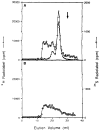
Fig. 1
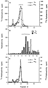
Fig. 1. Chromatography of proteolytic hydrolysates of lamins B and C on Sephadex LH-20
HeLa cells were labeled with [3H]MVA or [35S]cysteine, lamins B and C were isolated, solubilized in SDS, and extensively hydrolyzed with a combination of proteases, and the hydrolysates were concentrated on DEAE-Sephacel. 65% of the radioactivity in the hydrolysate of 3H-labeled lamin B adsorbed onto DEAE and could subsequently be eluted with formic acid/ethanol/water (2:8:1). In contrast, only 13 and 11%, respectively, of the radioactivity from the hydrolysates of 35S-labeled lamin B and lamin C behaved similarly. The figure shows chromatograms that were obtained when the DEAE-eluted, 3H- and 35S-labeled materials were each passed separately through Sephadex LH-20 in 20% formic acid in ethanol. Panel A, filled circles, 3H-labeled material from hydrolysate of lamin B (145,000 cpm loaded); open circles, corresponding, 35S-labeled material (100,000 cpm loaded). Panel B, 35S-labeled material from lamin C (80,000 cpm loaded). Note that an authentic standard of S-farnesyl cysteine coeluted with the major peak of 3H- label, shown in panel A, while cysteine and cystine emerged in the position indicated by the arrow. Hydrolysates of labeled lamin B yielded results like these in three different experiments, although the small peak of 3H-labeled material, that emerged at an elution volume of 20 ml, contained variable proportions (5–20%) of the total radioactivity. Hydrolysates of the [3H]MVA-labeled, minor nuclear protein of 66 kDa, described previously (5) also yielded results that were similar to those shown in panel A.
Fig. 2
Fig. 2. Reverse-phase and thin layer chromatography of 3H- and 35S-labeled proteolysis products of lamin B
Panel A, material corresponding to the major peak of radioactivity from each of the two chromatograms shown in Fig. 1A was passed through silica guard columns (not shown), then the 3H- and 35S-labeled samples were chromatographed separately by reverse-phase HPLC in methanol 0.1% trifluoroacetic acid (see “Experimental Procedures”). In the experiments shown 19,000 cpm of the 3H- and 5,300 cpm of the 35S-label were chromatographed separately to yield 10,000 and 2,000 cpm, respectively, in fractions 16 through 22. Note that pretreatment with the guard column removed 40% of the 35S radioactivity but only 20% of the 3H radioactivity, and that the guard column did not adsorb S-farnesyl cysteine. However, the guard column adsorbed more than 85% of the 35S radioactivity from the corresponding fraction of a lamin C hydrolysate, leaving insufficient radioactivity to be detected by reverse-phase HPLC. Panel B, fractions 16 through 22 from each of the chromatograms shown in panel A were pooled separately, concentrated, and further chromatographed by thin layer chromatography. In this experiment, 570 cpm of 3H- and 570 cpm of 35S-label was applied separately on adjacent lanes of a prescored thin layer chromatography plate and developed (see “Experimental Procedures”). The distribution of radioactivity in these two lanes is shown and was used to locate fractions of interest in two adjacent lanes, one loaded with 1,100 cpm of the 3H-label and the other with 1,100 cpm of the 35S-label. Fractions 2 and 17, respectively, corresponded to the origin and solvent front. Panel C, material corresponding to fractions 9 through 11 from the experiment shown in panel B was separately eluted from each of the two adjacent lanes described above, pooled, concentrated, and chromatographed by reverse-phase HPLC in methanol, 0.1% trifluoroacetic acid. In this experiment 340 cpm of the pooled 3H material and 500 cpm of the pooled 35S material were chromatographed separately to yield 220 and 370 cpm, respectively, in fractions 16 through 22. This represents 80 and 75%, respectively, of the total radioactivity actually recovered from the column.
Fig. 3
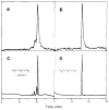
Fig. 3. Gas-liquid chromatographic analysis of [3H]MVA-derived material released upon treating lamin B with Raney nickel
3H-Labeled lamin B was solubilized with 8 M guanidine HCl, 0.2 M sodium phosphate, p H 7.0, and treated with Raney nickel. The released material was then extracted into pentane and analyzed by GLC before or after hydrogenation over platinum. Panel A, nonhydrogenated, 3H-labeled material released from lamin B by Raney nickel. Panel B, 3H-labeled material released from lamin B by Raney nickel that was subsequently hydrogenated over platinum. Similar results were obtained upon analyzing 3H-labeled material that had been released from the minor [3H]MVA-labeled protein of 66 kDa (5). Panel C, Raney nickel-released material from S-farnesyl cysteine. Panel D, material that had been similarly released from S-farnesyl cysteine, then hydrogenated over platinum before being analyzed. Authentic 2,6,10-trimethyldodecane (farnesane) co-chromatographed with this hydrogenated sample (not shown). Note that 58% of the 3H-labeled material that was released from lamin B was recovered from the column.
Fig. 4
Fig. 4. Enhanced electron ionization spectra of MVA-derived material released upon treating lamin B with Raney nickel
Lamin B from 1.2 ×10⁹ cells was solubilized with 8 M guanidine HCl, 0.2 M sodium phosphate, pH 7.0, then treated with Raney nickel. The released material was extracted into pentane and analyzed by GC/MS before or after hydrogenation. S-farnesyl cysteine was treated in a similar manner. Panel A, spectrum of a nonhydrogenated peak from the lamin B chromatogram which corresponded to the major peak of radioactivity in Fig. 3A. The tR of this spectrum was 18.93 min. The aliquot analyzed represents lamin B from 0.1 ×10⁹ cells. Panel B, spectrum of a hydrogenated peak from the lamin B chromatogram which corresponded to the peak of radioactivity in Fig. 3B. The tR of this spectrum was 14.43 min. The aliquot analyzed represents lamin B from 0.1× 10⁹ cells. Similar results were obtained upon the analysis of material that had been released from the minor protein of 66 kDa (5). Panel C, spectrum of the major peak from the Raney nickel-treated S-farnesyl cysteine chromatogram. The tR of this spectrum was 19.12 min. Panel D, spectrum of authentic 2,6,10-trimethyldodecane (farnesane). The tR of this spectrum was 14.52 min. All spectra shown have been enhanced to highlight ions of low abundance (see “Experimental Procedures”).
Fig. 5
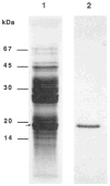
Fig. 5. Cyanogen bromide cleavage products of 3H-labeled lamin B
Purified 3H-labeled lamin B (~15 μg, 19,000 cpm) was treated with cyanogen bromide for 24 h in 70% formic acid. The cleavage products were separated on a 12.5% polyacrylamide gel, stained with Coomassie Blue (lane 1), and subsequently fluorographed (lane 2). The arrow indicates the labeled Coomassie band that was excised for amino acid sequence analysis. This 17-kDa peptide represented 50–100 pmol of material and contained 83% of the radioactivity recovered from the gel by direct radiometric analysis of excised gel slices after correction for background.
Fig. 6
Fig. 6. Alignment of the partial sequence of the labeled 17-kDa, cyanogen bromide cleavage product with the predicted carboxyl-terminal amino acid sequence of human lamin B
The 17-kDa labeled fragment (50–100 pmol) shown in Fig. 5, was transferred to a polyvinylidene difluoride membrane and partially sequenced. The resulting sequence of 18 amino acids (CNRr) was compared with the inferred sequence of human lamin B (HLb). Only the carboxyl 118 amino acids of lamin B are shown for comparison. The partial sequence mapped with near identity to the human lamin B sequence beginning at amino acid 482. The sequence mapped to this same position in the Xenopus LI sequence with an alignment score of 5.3 standard deviations. The conserved carboxyl Cys-A-A-X motif is underlined.

Similar articles

Cited by

References

    1. Schroepfer GJ. Annu Reu Biochem. 1981;50:585–621. - PubMed
    1. Schmidt RA, Schneider CJ, Glomset J. J Biol Chem. 1984;259:10175–10180. - PubMed
    1. Sinensky M, Logel J. Proc Natl Acad Sci U S A. 1985;82:3257–3261. - PMC - PubMed
    1. Maltese WA, Sheridan KM. J Cell Physiol. 1987;133:471–481. - PubMed
    1. Wolda SL, Glomset JA. J Biol Chem. 1988;263:5997–6000. - PubMed

Publication types

LinkOut - more resources