. 2016 Feb 5;12(2):e1005843.
doi: 10.1371/journal.pgen.1005843. eCollection 2016 Feb.

The Replisome-Coupled E3 Ubiquitin Ligase Rtt101Mms22 Counteracts Mrc1 Function to Tolerate Genotoxic Stress

Affiliations

The Replisome-Coupled E3 Ubiquitin Ligase Rtt101Mms22 Counteracts Mrc1 Function to Tolerate Genotoxic Stress

Raymond Buser et al. PLoS Genet. .

Abstract

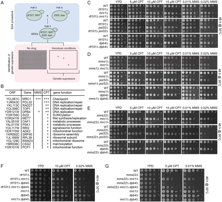
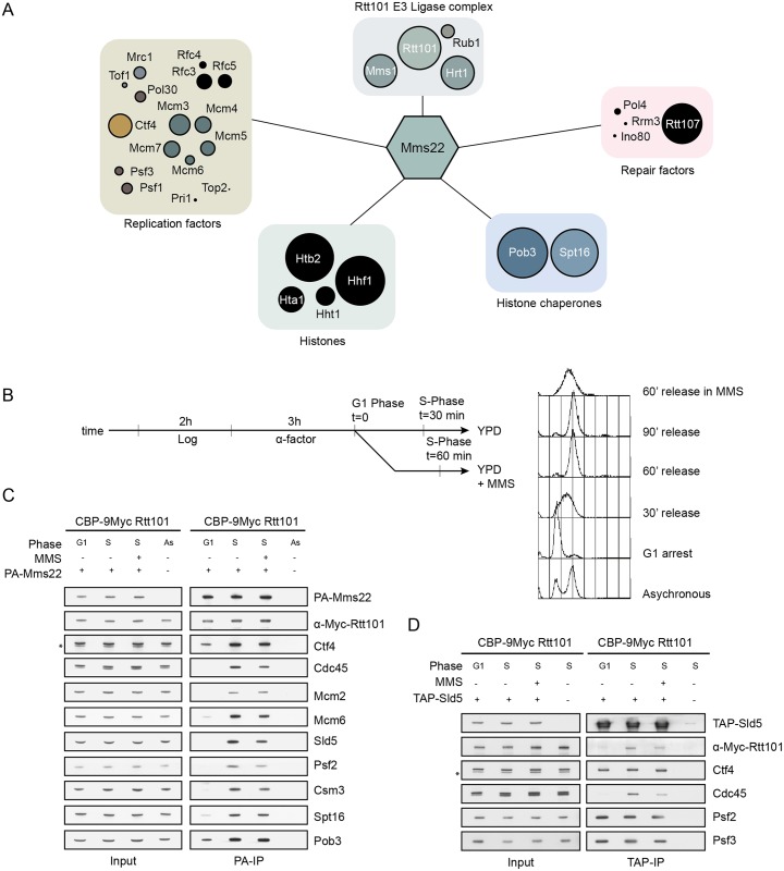
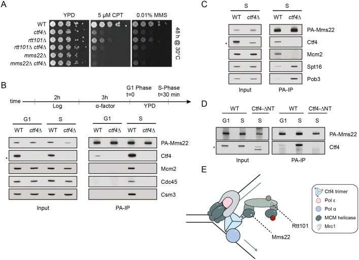
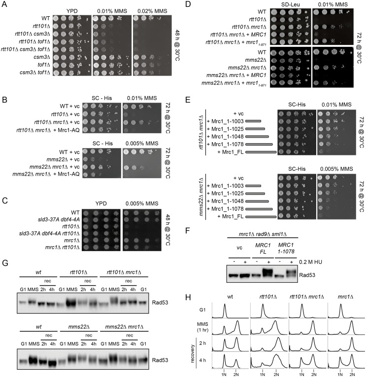
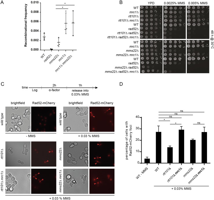
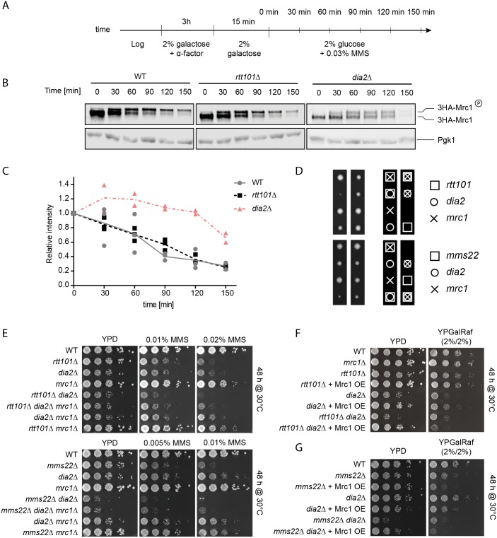
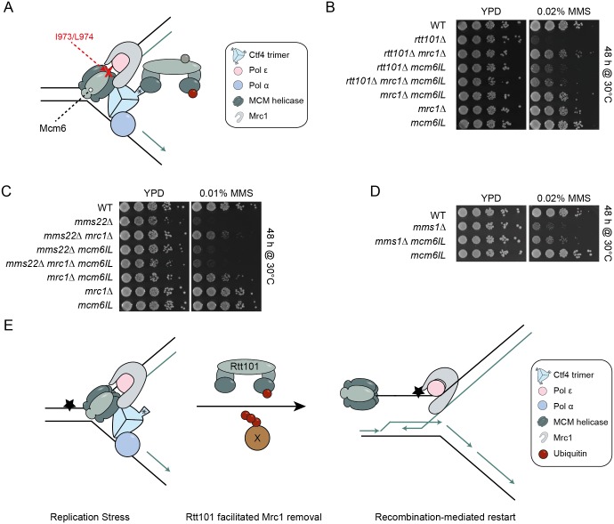
Faithful DNA replication and repair requires the activity of cullin 4-based E3 ubiquitin ligases (CRL4), but the underlying mechanisms remain poorly understood. The budding yeast Cul4 homologue, Rtt101, in complex with the linker Mms1 and the putative substrate adaptor Mms22 promotes progression of replication forks through damaged DNA. Here we characterized the interactome of Mms22 and found that the Rtt101(Mms22) ligase associates with the replisome progression complex during S-phase via the amino-terminal WD40 domain of Ctf4. Moreover, genetic screening for suppressors of the genotoxic sensitivity of rtt101Δ cells identified a cluster of replication proteins, among them a component of the fork protection complex, Mrc1. In contrast to rtt101Δ and mms22Δ cells, mrc1Δ rtt101Δ and mrc1Δ mms22Δ double mutants complete DNA replication upon replication stress by facilitating the repair/restart of stalled replication forks using a Rad52-dependent mechanism. Our results suggest that the Rtt101(Mms22) E3 ligase does not induce Mrc1 degradation, but specifically counteracts Mrc1's replicative function, possibly by modulating its interaction with the CMG (Cdc45-MCM-GINS) complex at stalled forks.


Conflict of interest statement

The authors have declared that no competing interests exist.

Figures

Fig 1
Fig 1. The Rtt101-Mms1-Mms22 E3 ubiquitin ligase genetically interacts with genes involved in DNA replication.
(A) and (B) Schematic representation of the synthetic genetic array (SGA) screening procedure (A), and table summarizing cellular functions of the main hits (B). Genotoxic conditions included MMS (0.01%) and CPT (5 μM) and colony size was qualitatively scored after 72 hours at 30°C. (+) weak suppression, (++) medium suppression, (+++) strong suppression. See Materials and Methods for a detailed description of the screening procedure and S1 Fig for an example of the raw data. (C) RTT101 genetically interacts with the replication genes MRC1, POL32 and DPB4. Serial dilution of wild-type (WT) or rtt101Δ, mrc1Δ, rtt101Δ mrc1Δ, pol32Δ, rtt101Δ pol32Δ, dpb4Δ and rtt101Δ dpb4Δ mutants were assayed on normal growth media (YPD), and media containing 5 μM, 10 μM and 15 μM CPT or 0.01% and 0.02% MMS. Cells were imaged after 48 hours of incubation at 30°C.(D) MMS1 phenocopies the genetic interactions of RTT101 with MRC1, POL32 and DPB4. Serial dilution of wild-type (WT) or mms1Δ, mrc1Δ, mms1Δ mrc1Δ, pol32Δ, mms1Δ pol32Δ, dpb4Δ and mms1Δ dpb4Δ mutants were assayed on normal growth media (YPD), and media containing 5 μM, 10 μM and 15 μM CPT or 0.01% and 0.02% MMS. Cells were imaged after 48 hours of incubation at 30°C. (E) MMS22 phenocopies the genetic interactions of RTT101 and MMS1 with MRC1 and DPB4, but not POL32. Serial dilution of wild-type (WT) or mms22Δ, mrc1Δ, mms22Δ mrc1Δ, pol32Δ, mms22Δ pol32Δ, dpb4Δ and mms22Δ dpb4Δ mutants were assayed on normal growth media (YPD), and media containing 5 μM, 10 μM and 15 μM CPT or 0.01% and 0.02% MMS. Cells were imaged after 48 hours of incubation at 30°C. (F) and (G). mrc1Δ and dpb4Δ rescue rtt101Δ and mms22Δ in an epistatic manner. Cells were spotted on the indicated media and imaged as described above in (C, D and E).
Fig 2
Fig 2. The substrate-specific adaptor Mms22 physically interacts with replisome components during S-phase.
(A) The S-phase specific Mms22 interaction network identified by MS/MS. PA-tagged Mms22 was immunoprecipitated from cells synchronized in S-phase and associated proteins were identified by MS-analysis. The hits were grouped according to known functions and the ring diameter is proportional to the percent of coverage measured for the indicated proteins. (B) and (C) Mms22 physically interacts with components of the replisome during S-phase. Cells endogenously expressing CBP-9Myc-Rtt101 were transformed with PA-Mms22 and synchronized in S-phase by release from α-factor arrest as schematically outlined (B), and either treated or not with 0.03% MMS to induce fork stalling. Visualizing the DNA content by flow cytometry monitored cell synchronization. PA-Mms22 was immunoprecipitated from the indicated cell extracts and associated proteins detected by immunoblotting using specific antibodies (C). The asterisk (*) indicates an unspecific band. (D) S-phase specific interaction of Rtt101 with replisome components. Cells endogenously expressing CBP-9Myc-Rtt101 and TAP-Sld5 were synchronized and treated as in (C). TAP-Sld5 was purified from the indicated extracts and interacting proteins visualized by immunoblotting with specific antibodies.
Fig 3
Fig 3. The WD40 domain of Ctf4 recruits the Rtt101Mms22 E3 ubiquitin ligase to the replisome progression complex.
(A) CTF4 is epistatic with RTT101 and MMS22. Exponentially growing wild-type (WT), ctf4Δ, rtt101Δ, mms22Δ, rtt101Δ ctf4Δ and mms22Δ ctf4Δ cells were spotted in serial dilution on normal growth media (YPD) and media containing 5 μM CPT or 0.01% MMS. The plates were imaged after 48 hours incubation at 30°C. (B) and (C) Ctf4 tethers the Rtt101 E3 ligase to the replisome. Wild-type (WT) and ctf4Δ cells were transformed with a plasmid expressing PA-Mms22, arrested in α-factor (G1) or synchronously released into S-phase (S) (diagram). PA-Mms22 was purified from G1 or S-phase extracts and interacting replisome (B) or FACT (C) components were detected using specific antibodies. The asterisk (*) indicates an unspecific band. (D) The WD40 domain of Ctf4 is crucial for Mms22 interaction with the RPC. Wild-type (WT) and ctf4-ΔNT cells were synchronized as described in (B). PA-Mms22 was purified from extracts prepared from cells in G1 or S-phase and its interaction with Ctf4 was monitored by immunoblotting with specific antibodies. (E) Model of the Rtt101Mms22 E3 ubiquitin ligase interacting with the replisome. Ctf4 together with Mrc1 connects the DNA polymerases to the MCM helicase at replication forks. Mms22 interacts with the amino-terminal domain of Ctf4 (*), which is required to recruit the Rtt101Mms22 E3 ubiquitin ligase to active forks.
Fig 4
Fig 4. The replicative function of Mrc1 compensates for replication defects in cells lacking components of the Rtt101Mms22 E3 ubiquitin ligase.
(A) Suppression of the growth phenotype of cells lacking components of the Rtt101Mms22 E3 ligase is specific to Mrc1. Serial dilution of wild-type (WT), rtt101Δ, csm3Δ, csm3Δ rtt101Δ, tof1Δ, tof1Δ rtt101Δ, tof1Δ rtt101Δ csm3Δ and csm3Δ tof1Δ cells were analyzed on normal growth media (YPD) with or without 0.01% and 0.02% MMS.(B) The checkpoint-defective Mrc1-AQ allele does not suppress the sensitivity of mms22Δ or rtt101Δ cells to MMS. Serial dilution of wild-type (WT), rtt101Δ, mrc1Δ, mms22Δ, rtt101Δ mrc1Δ, mms22Δ mrc1Δ strains were assayed on selective growth media (-His) with or without MMS. The lack of MRC1 was complemented as indicated with plasmids expressing either vector control (vc) or Mrc1-AQ. Plates were imaged after 72 hours incubation at 30°C. (C) De-repressing late origins is not sufficient to suppress rtt101Δ MMS sensitivity. Strains with the indicated genotypes were serial diluted on the indicated media as described above. Plates were imaged after 48 hours incubation at 30°C. (D)-(F) C-terminal truncations of Mrc1 can provide a genetic rescue of rtt101Δ and mms22Δ cells. (D and E) Cells expressing either a vector control (vc), wild-type (FL = full length) or the indicated Mrc1 truncation mutants were spotted on selective growth media (-His) with or without MMS. The black bars represent the c-terminally truncated Mrc1 proteins. Plates were imaged after 72 hours incubation at 30°C. (F) mrc1Δ rad9Δ sml1Δ cells were transformed with an empty control vector (vc), or plasmids encoding either wild-type (MRC1FL) or the MRC11-1078 truncation allele and synchronously released into media with (+) or without (-) 0.2M hydroxyurea (HU). Rad53 phosphorylation was analyzed by immunoblotting of protein extracts with anti-Rad53 antibody. (G) and (H) The checkpoint defect of rtt101Δ and mms22Δ cells is alleviated by loss of MRC1. Cells were synchronized in G1 phase using α-factor and released into media containing 0.01% MMS for 60 min. Stalled forks were then allowed to restart in normal growth media (recovery) and the checkpoint status was monitored by Rad53 phosphorylation (G) and flow cytometry (H) at the indicated time points (in hours (h); rec = recovery).
Fig 5
Fig 5. Mrc1 prevents Rad52-mediated HR in mms22Δ and rtt101Δ cells.
(A) Deleting MRC1 compensates for the HR defect of mms22Δ cells. Wild-type (WT) and the indicated mutant strains were transformed with a homologous recombination reporter plasmid (YCpHR), and the recombination frequency (%) was quantified from three independent experiments as schematically outlined in S6 Fig on plates containing 60 μg/ml canavanine. A one-way ANOVA analysis was used for the statistical analysis. * represents a significant difference with 95% confidence. Error bars indicate standard deviation. (B) The growth restoration of rtt101Δ mrc1Δ and mms22Δ mrc1Δ cells on MMS is RAD52-dependent. Serial dilution of wild-type (WT) or mrc1Δ, rtt101Δ, rtt101Δ mrc1Δ, rtt101Δ mrc1Δ rad52Δ, mms22Δ, mms22Δ mrc1Δ, mms22Δ mrc1Δ rad52Δ mutants were assayed on normal growth media and media containing 0.0025% or 0.005% MMS and imaged after 48 hours incubation at 30°C. (C) and (D) rtt101Δ and mms22Δ cells are defective in forming RAD52 foci. Synchronized cells were released into the presence of MMS as depicted (top diagram) and cells with Rad52-mCherry foci were scored (C). The experiment was performed with two biological replicates, and for each genotype a minimum of 400 cells per replicate were counted (D). * represents a significant difference with 95% confidence intervals following a one-way ANOVA analysis. ns: not significant (below 95% confidence). Error bars indicate standard deviation.
Fig 6
Fig 6. Rtt101Mms22 and SCFDia2 counteract Mrc1 activity by distinct mechanisms. (A)–(C) Mrc1 stability is not altered in cells lacking RTT101.
Wild-type (WT), rtt101Δ or dia2Δ cells expressing 3HA-tagged Mrc1 from the inducible GAL1,10-promoter were synchronized in G1 phase using α-factor in 2% galactose and released into S-phase in 2% galactose as outlined in (A). Subsequently, 0.03% MMS and 2% glucose was added to induce fork stalling and repress HA-Mrc1 expression, respectively. Samples were collected at the indicated time points (min) (A), quantified by anti-HA immunoblotting (B) and shown as scatter plots of individual biological replicates with their means (C). Immunoblotting for Pgk1 controls for equal loading. The position of phosphorylated (3HA-Mrc1-P) and unphosphorylated 3HA-Mrc1 is indicated. (D) RTT101 and MMS22 are synthetic-sick with DIA2. Tetrad analysis from sporulated heterozygote rtt101Δ dia2Δ mrc1Δ and mms22Δ dia2Δ mrc1Δ diploids. Boxes (□), crosses (X) and circles (O) indicate that the haploid cells lack RTT101/MMS22, MRC1 or DIA2, respectively. Note that rtt101Δ dia2Δ and mms22Δ dia2Δ double mutants indicated by boxes and circles are barely viable. (E) rtt101Δ dia2Δ and mms22Δ dia2Δ double mutants can be rescued by deletion of MRC1. Serial dilution of the indicated strains obtained from the tetrad dissections shown in (D) were assayed on normal growth media (YPD) and media containing 0.02%, 0.01% or 0.005% MMS. The plates were imaged after 48 hours of incubation at 30°C. (F) and (G) Overexpression of Mrc1 is lethal for rtt101Δ dia2Δ (F) and mms22Δ dia2Δ (G) double mutants. Serial dilution of wild-type (WT) and the indicated mutant strains overexpressing Mrc1 from the galactose-inducible GAL1,10-promoter were assayed on normal growth media containing 2% glucose (GAL1,10-promoter off), 2% galactose/2% raffinose (GAL1,10-promoter on). The plates were imaged after 48 hours of incubation at 30°C.
Fig 7
Fig 7. Altered regulation of replicative polymerases and the CMG may allow HR mediated fork restart in cells lacking Rtt101Mms22 activity.
(A) mcm6-IL reduces its affinity to physically interact with Mrc1. Mutation of the indicated amino acids perturbs the interaction between Mcm6 and Mrc1 and is reported to promote uncoupling of the leading strand polymerase from the MCM replicative helicase [14]. The asterisk (*) indicates the WD40 domain of Ctf4. (B)-(D) The mcm6-IL allele rescues the genotoxic sensitivity of rtt101Δ (B), mms1Δ (C) or mms22Δ (D) mutants and is epistatic with MRC1. Cells with the indicated genotypes were spotted onto either YPD or YPD plates containing 0.02% or 0.01% MMS. Images were taken after 48 hours of incubation at 30°C. (E) Model of Rtt101Mms22-facilitated removal of Mrc1 to allow HR-mediated repair/restart of stalled replication forks. The Rtt101Mms22 E3 ligase associates with replisomes by binding to Ctf4. When a replication fork encounters an obstacle (star), our data suggest that Rtt101Mms22 ubiquitylates a so far unidentified factor (X), which modulates the interaction between Mrc1 and the MCM helicase (Mcm6 is in light grey). This remodeling results in the Rad52-mediated repair/restart of the stressed replisome in order to by-pass the obstacle.

Similar articles

Cited by

References

    1. Hodgson B, Calzada A, Labib K (2007) Mrc1 and Tof1 regulate DNA replication forks in different ways during normal S phase. Mol Biol Cell 18: 3894–3902. 10.1091/mbc.E07-05-0500 - DOI - PMC - PubMed
    1. Gambus A, Jones RC, Sanchez-Diaz A, Kanemaki M, van Deursen F, et al. (2006) GINS maintains association of Cdc45 with MCM in replisome progression complexes at eukaryotic DNA replication forks. Nat Cell Biol 8: 358–366. 10.1038/ncb1382 - DOI - PubMed
    1. Gambus A, van Deursen F, Polychronopoulos D, Foltman M, Jones RC, et al. (2009) A key role for Ctf4 in coupling the MCM2-7 helicase to DNA polymerase alpha within the eukaryotic replisome. EMBO J 28: 2992–3004. 10.1038/emboj.2009.226 - DOI - PMC - PubMed
    1. Tanaka H, Katou Y, Yagura M, Saitoh K, Itoh T, et al. (2009) Ctf4 coordinates the progression of helicase and DNA polymerase alpha. Genes Cells 14: 807–820. 10.1111/j.1365-2443.2009.01310.x - DOI - PubMed
    1. Simon AC, Zhou JC, Perera RL, van Deursen F, Evrin C, et al. (2014) A Ctf4 trimer couples the CMG helicase to DNA polymerase α in the eukaryotic replisome. Nature 510: 293–297. 10.1038/nature13234 - DOI - PMC - PubMed

Publication types

MeSH terms