Review
. 2016 Jun:54:188-97.
doi: 10.1016/j.semcdb.2016.01.042. Epub 2016 Feb 3.

Repression of harmful meiotic recombination in centromeric regions

Affiliations
Review

Repression of harmful meiotic recombination in centromeric regions

Mridula Nambiar et al. Semin Cell Dev Biol. 2016 Jun.

Abstract

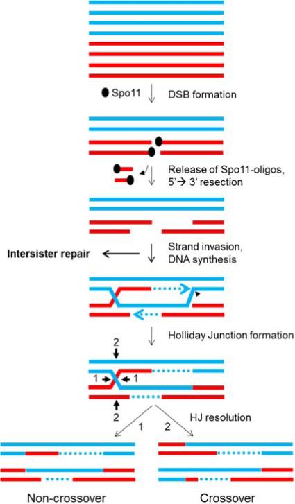
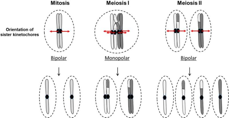
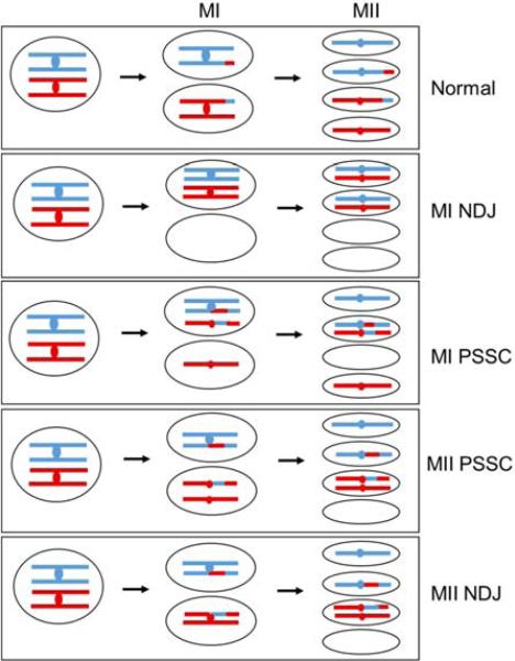
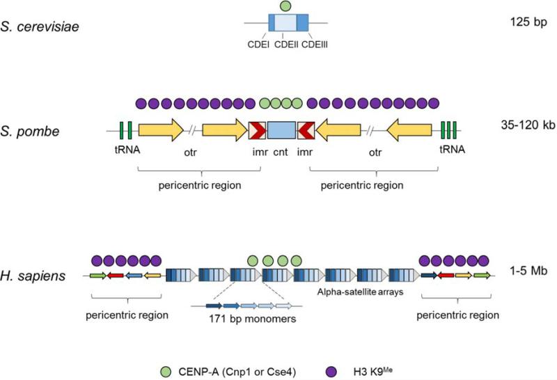
During the first division of meiosis, segregation of homologous chromosomes reduces the chromosome number by half. In most species, sister chromatid cohesion and reciprocal recombination (crossing-over) between homologous chromosomes are essential to provide tension to signal proper chromosome segregation during the first meiotic division. Crossovers are not distributed uniformly throughout the genome and are repressed at and near the centromeres. Rare crossovers that occur too near or in the centromere interfere with proper segregation and can give rise to aneuploid progeny, which can be severely defective or inviable. We review here how crossing-over occurs and how it is prevented in and around the centromeres. Molecular mechanisms of centromeric repression are only now being elucidated. However, rapid advances in understanding crossing-over, chromosome structure, and centromere functions promise to explain how potentially deleterious crossovers are avoided in certain chromosomal regions while allowing beneficial crossovers in others.

Keywords: Aneuploidy; Centromeres; Chromosome segregation; Crossing-over; Homologous recombination; Meiosis.


Figures

Figure 1
Figure 1. Formation of crossovers and non-crossovers at Spo11-induced DSBs
Red and blue lines are single DNA strands, and each pair depicts one sister chromatid of each homologous chromosome (the non-interacting sisters are shown only in the top panel); dotted lines are DNA synthesized during DSB repair. After meiotic replication, Spo11 or its ortholog induces DSBs and covalently attaches to the 5’ ends. Spol11 is then cleaved from the ends by an endonuclease to release a short oligonucleotide attached to Spo11. The 5’ ends are further resected to generate long 3’ overhangs, which invade intact duplex DNA, either the homolog or the sister chromatid; this side reaction with the sister produces no genetic recombinants. Holliday junctions, either single (shown) or double (not shown), are formed and then resolved into either crossovers or non-crossovers (respectively with the non-parental or parental DNA configuration flanking the region of DNA exchange). These reactions have been demonstrated by direct DNA analysis in budding and fission yeasts and are inferred, from genetic and cytological data and limited DNA analysis, to occur in other species. In an alternative to Holliday junction formation, strand invasion at the third step can prime limited DNA synthesis; the unwound product can anneal with the other initially broken DNA end and produce a non-crossover. This proposed reaction is called synthesis-dependent strand-annealing.
Figure 2
Figure 2. Orientation of kinetochores during mitosis and meiosis
In mitosis, kinetochores attach to the centromeres of the sister chromatids, orient towards opposite poles (bipolar orientation), and segregate into separate daughter nuclei. During the first meiotic division, however, both sister kinetochores orient towards the same pole (monopolar orientation) and segregate into the same daughter nucleus. Proper direction of segregation is ensured by sister chromatid cohesion and the tension generated due to crossovers (light grey-dark grey junction) between homologs. This reductional division results in separation of the homologous centromeres and the attached chromosomal arms and reduces the number of chromosomes by half. The second meiotic division is similar to mitosis, and bipolar orientation of the sister kinetochores results in separation of the chromatids into four haploid nuclei.
Figure 3
Figure 3. Meiotic chromosomal missegregation resulting from pericentric crossovers
Red and blue lines depict homologs (duplex DNA); red and blue dots depict centromeres. Crossovers, depicted by red-blue junctions, in chromosomal arms are essential for proper segregation of chromosomes during meiosis, but crossovers too near the centromere are harmful. Normally during MI, the centromeres of homologs with their attached chromosomal arms segregate to opposite poles, whereas in MII the sister centromeres segregate to opposite poles, giving rise to four haploid nuclei. During non-disjunction (NDJ) of homologs in MI, which primarily arises due to lack of crossovers in the arms, both homologs migrate to the same pole and then segregate properly at MII, giving rise to two nullisomes (nuclei lacking a chromosome) and two disomes (nuclei with two chromosome copies). Precocious separation of sister chromatids (PSSC) occurs when cohesion is lost between the sister chromatids and can occur in either MI or MII. Crossovers too close to the centromeres (pericentric crossovers) are associated with such PSSC events. In MI PSSC, sister centromeres of one homolog segregate to opposite poles at MI followed by proper MII, giving rise to a nullisome and a disome containing homologous centromeres. In MII PSSC, sisters stay in the same nucleus at MI but missegregate at MII, giving rise to a nullisome and a disome containing sister centromeres. MII NDJ has proper MI but aberrant segregation at MII, resulting in a fate similar to MII PSSC. These aberrant events are also linked to the presence of pericentric crossovers. Some of these missegregation events give rise to similar types of aneuploids, but careful tetrad analysis with multiple markers can distinguish them.
Figure 4
Figure 4. Structure of centromeres in yeasts and humans
The budding yeast S. cerevisiae has 125 bp (“point”) centromeres with three conserved regions – CDEI, II and III. A single nucleosome containing the centromere-specific histone H3 variant Cse4 (CENP-A in most other organisms) occupies the whole centromere. The fission yeast S. pombe contains large 35 – 120 kb (“regional”) centromeres consisting of a central core (cnt) surrounded by innermost repeats (imr) and outermost repeats (otr). The central core contains nucleosomes with CENP-A (Cnp1 in S. pombe), while the inverted repeats bear H3 K9me histones that form the pericentric heterochromatin. Human centromeres have large 1 – 5 Mb arrays of repetitive DNA consisting of 171 bp alpha-satellite repeats as the basic repeating unit. An array consists of multiple copies of higher order repeats which themselves are made up of multiple alpha-satellites that are diverged from each other. These arrays contain CENP-A interspersed with H3 K4me histones. The pericentric region contains stretches of monomeric alpha-satellite repeats that are in random orientation and are bound by H3 K9me histones that form the heterochromatin. The complete organization of human centromeres is still being worked out, and the schematic shown here is a model based on current understanding.

Similar articles

Cited by

References

    1. Przewloka MR, Glover DM. The kinetochore and the centromere: a working long distance relationship. Annu Rev Genet. 2009;43:439–465. - PubMed
    1. Nagaoka SI, Hassold TJ, Hunt PA. Human aneuploidy: mechanisms and new insights into an age old problem. Nat Rev Genet. 2012;13:493–504. - PMC - PubMed
    1. Lam I, Keeney S. Mechanism and regulation of meiotic recombination initiation. Cold Spring Harb Perspect Biol. 2015;7:a016634. - PMC - PubMed
    1. Keeney S. Spo11 and the formation of DNA double-strand breaks in meiosis. In: Egel R, Lankenau D-H, editors. Recombination and meiosis: crossing-over and disjunction. Springer-Verlag; Berlin: 2007. pp. 81–123.
    1. Smith KN, Nicolas A. Recombination at work for meiosis. Current Opinion in Genetics and Development. 1998;8:200–211. - PubMed

Publication types

LinkOut - more resources