Pedicle screw fixation and posterior fusion for lumbar degenerative diseases: effects on individual paraspinal muscles and lower back pain; a single-center, prospective study
- PMID: 26850001
- PMCID: PMC4744382
- DOI: 10.1186/s12891-016-0927-9
Pedicle screw fixation and posterior fusion for lumbar degenerative diseases: effects on individual paraspinal muscles and lower back pain; a single-center, prospective study
Abstract
Background: To the best of our knowledge, there have been no reports on the points at which the denervated multifidus and erector spinae muscles become reinnervated after pedicle screw fixation and posterior fusion in patients with lumbar degenerative diseases. Our study was designed to confirm reinnervation of denervated paraspinal muscles following pedicle screw fixation and posterior fusion and to confirm alleviation of the patients' lower back pain (LBP).
Methods: In this prospective study, we enrolled 67 patients who had undergone pedicle screw fixation and posterior fusion. The surgery had alleviated their leg pain, but the patients complained of LBP at the L3-5 level 3 months after the surgery. The patients were divided into two groups (I and II) according to the level at which pain was experienced. Paraspinal mapping scores were recorded preoperatively and 3, 6, 12, and 18 months postoperatively. Oswestry Disability Index and visual analogue scale scores were determined. Regression analyses using a general linear model and a mixed model were performed.
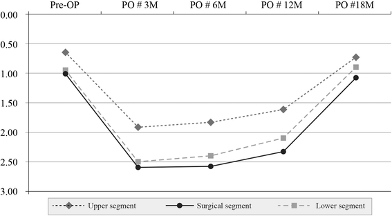
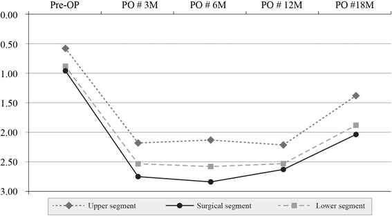
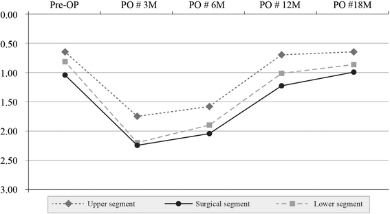
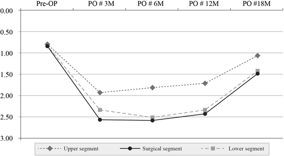
Results: Pedicle screw fixation and posterior fusion significantly denervated the multifidus and erector spinae not only in the surgical segment, but also in adjacent segments. Group I patients displayed reinnervation in the denervated erector spinae and multifidus muscles at 12 and 18 months, respectively. In contrast, group II showed reinnervation only in of the denervated erector spinae of the upper segment at 18 months, with no other areas of reinnervation. Postoperative LBP was significantly diminished at 12 months in group I and at 18 months in group II. There was also significantly less LBP at 6 months (prior to reinnervation of the paraspinal muscles).
Conclusions: The denervated multifidus and erector spinae muscles at L4-5, which had been denervated using pedicle screw fixation and posterior fusion, were significantly reinnervated at 18 months postoperatively, whereas patients with denervation at L3-5 had only a tendency to be reinnervated at follow-up. Postoperative LBP in these patients was significantly diminished at the follow-up visits.
Figures
Similar articles
-
The recovery of damaged paraspinal muscles by posterior surgical treatment for patients with lumbar degenerative diseases and its clinical consequence.J Back Musculoskelet Rehabil. 2017;30(4):801-809. doi: 10.3233/BMR-150455. J Back Musculoskelet Rehabil. 2017. PMID: 28372312
-
Quantitative analysis of paraspinal muscle atrophy after oblique lateral interbody fusion alone vs. combined with percutaneous pedicle screw fixation in patients with spondylolisthesis.BMC Musculoskelet Disord. 2020 Jan 14;21(1):30. doi: 10.1186/s12891-020-3051-9. BMC Musculoskelet Disord. 2020. PMID: 31937277 Free PMC article.
-
The Effect of Paraspinal Muscle Degeneration on Distal Pedicle Screw Loosening Following Corrective Surgery for Degenerative Lumbar Scoliosis.Spine (Phila Pa 1976). 2020 May 1;45(9):590-598. doi: 10.1097/BRS.0000000000003336. Spine (Phila Pa 1976). 2020. PMID: 31770334
-
Paraspinal muscle changes after single-level posterior lumbar fusion: volumetric analyses and literature review.BMC Musculoskelet Disord. 2020 Feb 5;21(1):73. doi: 10.1186/s12891-020-3104-0. BMC Musculoskelet Disord. 2020. PMID: 32024500 Free PMC article. Review.
-
Does pre-operative multifidus morphology on MRI predict clinical outcomes in adults following surgical treatment for degenerative lumbar spine disease? A systematic review.Eur Spine J. 2020 Jun;29(6):1318-1327. doi: 10.1007/s00586-020-06423-6. Epub 2020 Apr 23. Eur Spine J. 2020. PMID: 32328791
Cited by
-
Use of a life-size three-dimensional-printed spine model for pedicle screw instrumentation training.J Orthop Surg Res. 2018 Apr 16;13(1):86. doi: 10.1186/s13018-018-0788-z. J Orthop Surg Res. 2018. PMID: 29661210 Free PMC article.
-
Reattachment of the Multifidus Tendon in Lumbar Surgery to Decrease Postoperative Back Pain: A Technical Note.Cureus. 2019 Dec 12;11(12):e6366. doi: 10.7759/cureus.6366. Cureus. 2019. PMID: 31938648 Free PMC article.
-
Muscular changes after minimally invasive versus open spinal stabilization of thoracolumbar fractures: A literature review.J Musculoskelet Neuronal Interact. 2018 Mar 1;18(1):62-70. J Musculoskelet Neuronal Interact. 2018. PMID: 29504580 Free PMC article. Review.
-
Percutaneous Sacroplasty for Symptomatic Sacral Pedicle Screw Loosening.Indian J Orthop. 2022 Nov 22;57(1):96-101. doi: 10.1007/s43465-022-00773-7. eCollection 2023 Jan. Indian J Orthop. 2022. PMID: 36660492 Free PMC article.
-
Ti-24Nb-4Zr-8Sn Alloy Pedicle Screw Improves Internal Vertebral Fixation by Reducing Stress-Shielding Effects in a Porcine Model.Biomed Res Int. 2018 Feb 8;2018:8639648. doi: 10.1155/2018/8639648. eCollection 2018. Biomed Res Int. 2018. PMID: 29581988 Free PMC article.
References
-
- Helenius I, Remes V, Yrjönen T, Ylikoski M, Schlenzka D, Helenius M, et al. Comparison of long-term functional and radiologic outcomes after Harrington instrumentation and spondylodesis in adolescent idiopathic scoliosis: a review of 78 patients. Spine (Phila Pa 1976) 2002;27:176–80. doi: 10.1097/00007632-200201150-00010. - DOI - PubMed
Publication types
MeSH terms
LinkOut - more resources
Full Text Sources
Other Literature Sources
Medical
Miscellaneous

