

Increasing the strength and bioactivity of collagen scaffolds using customizable arrays of 3D-printed polymer fibers
- PMID: 26850145
- PMCID: PMC5030103
- DOI: 10.1016/j.actbio.2016.02.004
Increasing the strength and bioactivity of collagen scaffolds using customizable arrays of 3D-printed polymer fibers
Abstract
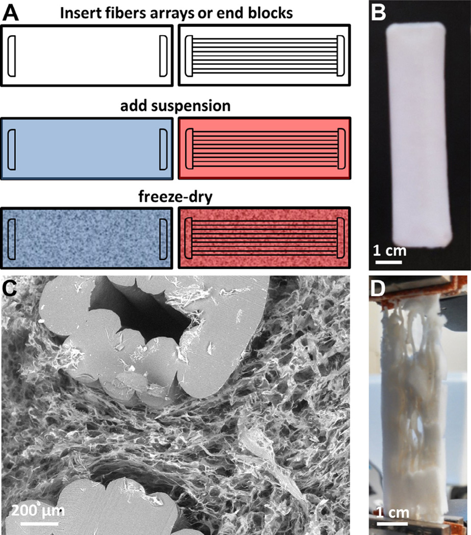
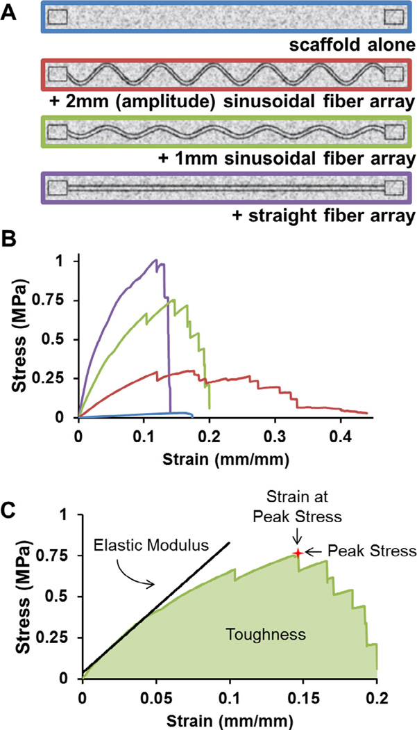
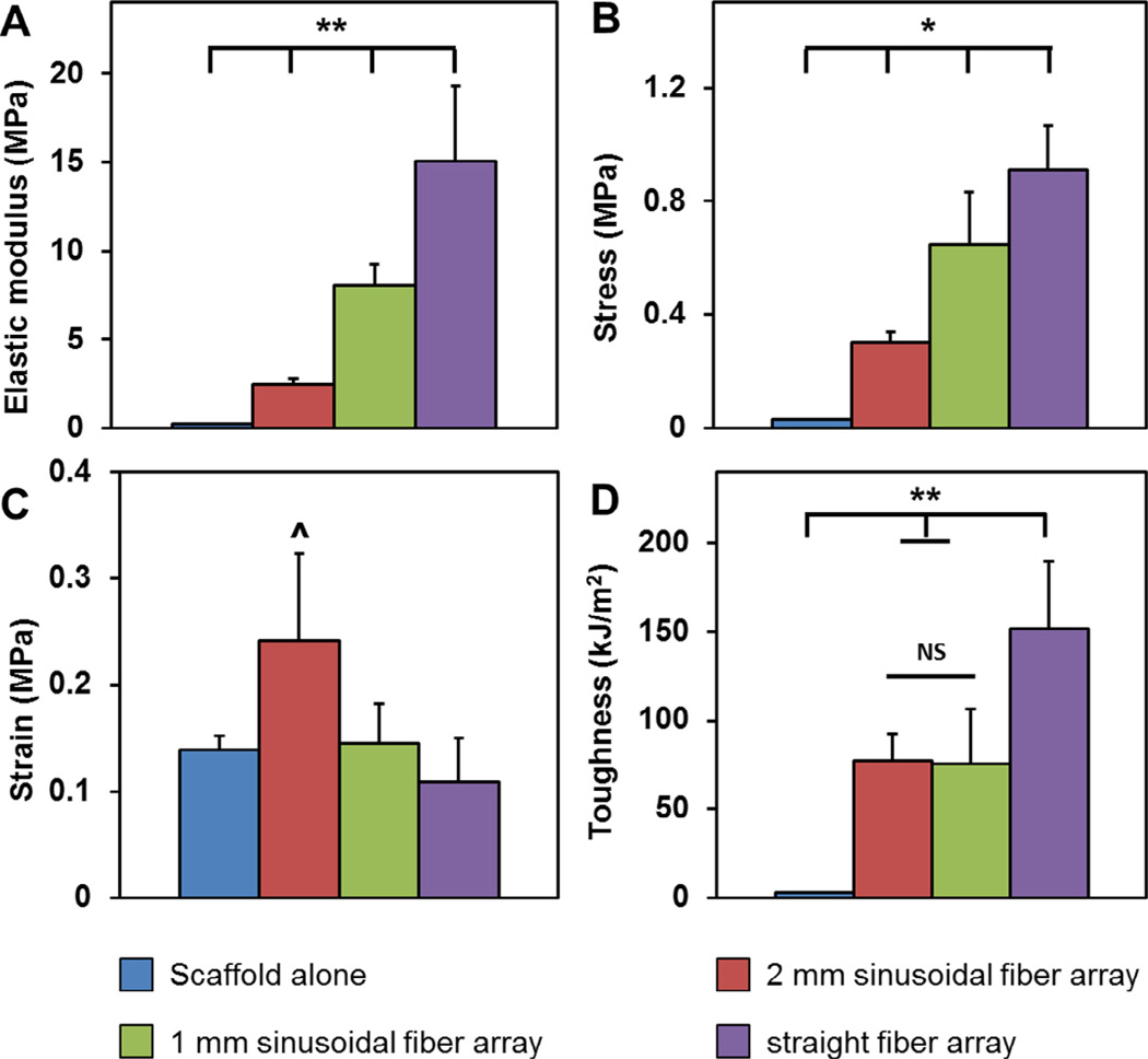
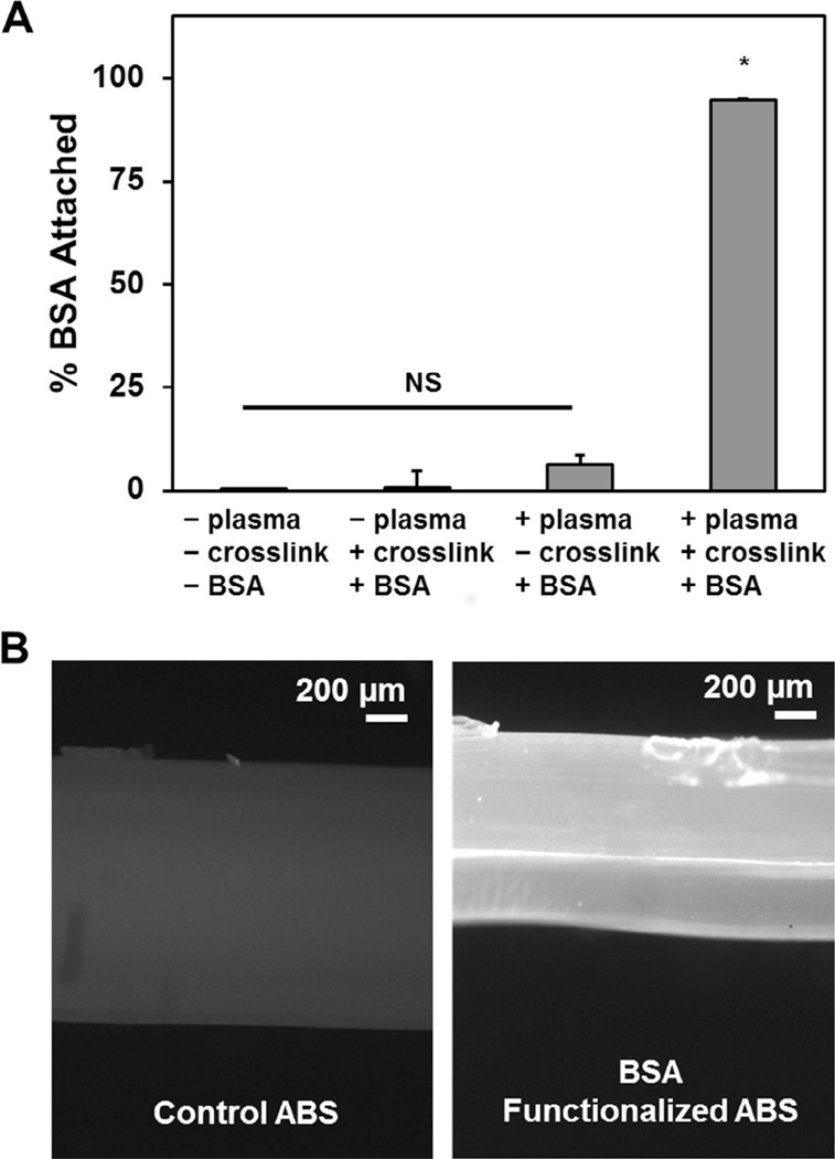
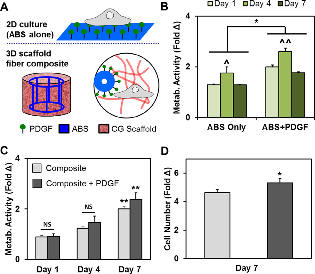
Tendon is a highly aligned connective tissue which transmits force from muscle to bone. Each year, people in the US sustain more than 32 million tendon injuries. To mitigate poor functional outcomes due to scar formation, current surgical techniques rely heavily on autografts. Biomaterial platforms and tissue engineering methods offer an alternative approach to address these injuries. Scaffolds incorporating aligned structural features can promote expansion of adult tenocytes and mesenchymal stem cells capable of tenogenic differentiation. However, appropriate balance between scaffold bioactivity and mechanical strength of these constructs remains challenging. The high porosity required to facilitate cell infiltration, nutrient and oxygen biotransport within three-dimensional constructs typically results in insufficient biomechanical strength. Here we describe the use of three-dimensional printing techniques to create customizable arrays of acrylonitrile butadiene styrene (ABS) fibers that can be incorporated into a collagen scaffold under development for tendon repair. Notably, mechanical performance of scaffold-fiber composites (elastic modulus, peak stress, strain at peak stress, and toughness) can be selectively manipulated by varying fiber-reinforcement geometry without affecting the native bioactivity of the collagen scaffold. Further, we report an approach to functionalize ABS fibers with activity-inducing growth factors via sequential oxygen plasma and carbodiimide crosslinking treatments. Together, we report an adaptable approach to control both mechanical strength and presence of biomolecular cues in a manner orthogonal to the architecture of the collagen scaffold itself.
Statement of significance: Tendon injuries account for more than 32 million injuries each year in the US alone. Current techniques use allografts to mitigate poor functional outcomes, but are not ideal platforms to induce functional regeneration following injury. Tissue engineering approaches using biomaterial substrates have significant potential for addressing these defects. However, the high porosity required to facilitate cell infiltration and nutrient transport often dictates that the resultant biomaterials has insufficient biomechanical strength. Here we describe the use of three-dimensional printing techniques to generate customizable fiber arrays from ABS polymer that can be incorporated into a collagen scaffold under development for tendon repair applications. Notably, the mechanical performance of the fiber-scaffold composite can be defined by the fiber array independent of the bioactivity of the collagen scaffold design. Further, the fiber array provides a substrate for growth factor delivery to aid healing.
Keywords: Composite CG scaffolds; Tendon tissue engineering; Three-dimensional printing; Tunable mechanics.
Copyright © 2016 Acta Materialia Inc. Published by Elsevier Ltd. All rights reserved.
Figures
References
-
- James R, Kesturu G, Balian G, Chhabra AB. Tendon: biology, biomechanics, repair, growth factors, and evolving treatment options. J. Hand Surg. Am. 2008;33:102–112. - PubMed
-
- Liu Y, Ramanath HS, Wang DA. Tendon tissue engineering using scaffold enhancing strategies. Trends Biotechnol. 2008;26:201–209. - PubMed
-
- O’Brien FJ, Harley BA, Yannas IV, Gibson LJ. The effect of pore size on cell adhesion in collagen-GAG scaffolds. Biomaterials. 2005;26:433–441. - PubMed
-
- O’Brien FJ, Harley BA, Waller MA, Yannas IV, Gibson LJ, Prendergast PJ. The effect of pore size on permeability and cell attachment in collagen scaffolds for tissue engineering. Technol. Health Care. 2007;15:3–17. - PubMed
Publication types
MeSH terms
Substances
Grants and funding
LinkOut - more resources
Full Text Sources
Other Literature Sources
Research Materials
