. 2016 Apr;170(4):2136-45.
doi: 10.1104/pp.15.01733. Epub 2016 Feb 5.

Auxin-Independent NAC Pathway Acts in Response to Explant-Specific Wounding and Promotes Root Tip Emergence during de Novo Root Organogenesis in Arabidopsis

Affiliations

Auxin-Independent NAC Pathway Acts in Response to Explant-Specific Wounding and Promotes Root Tip Emergence during de Novo Root Organogenesis in Arabidopsis

Xiaodong Chen et al. Plant Physiol. 2016 Apr.

Abstract

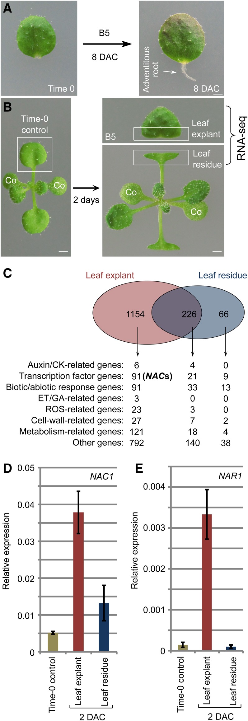
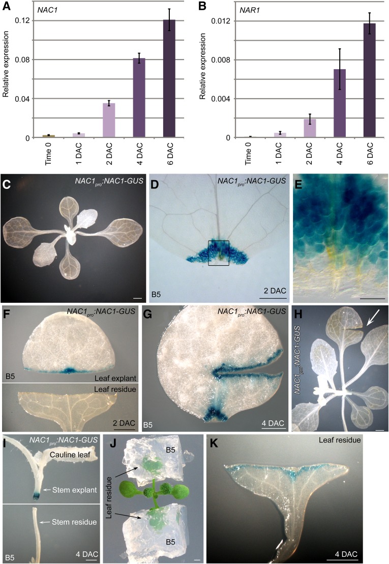
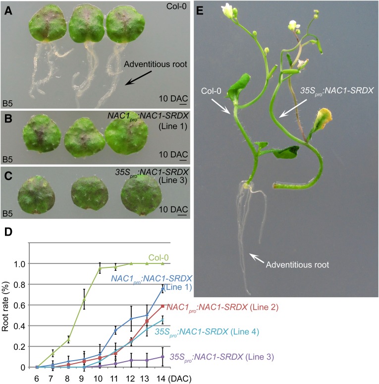
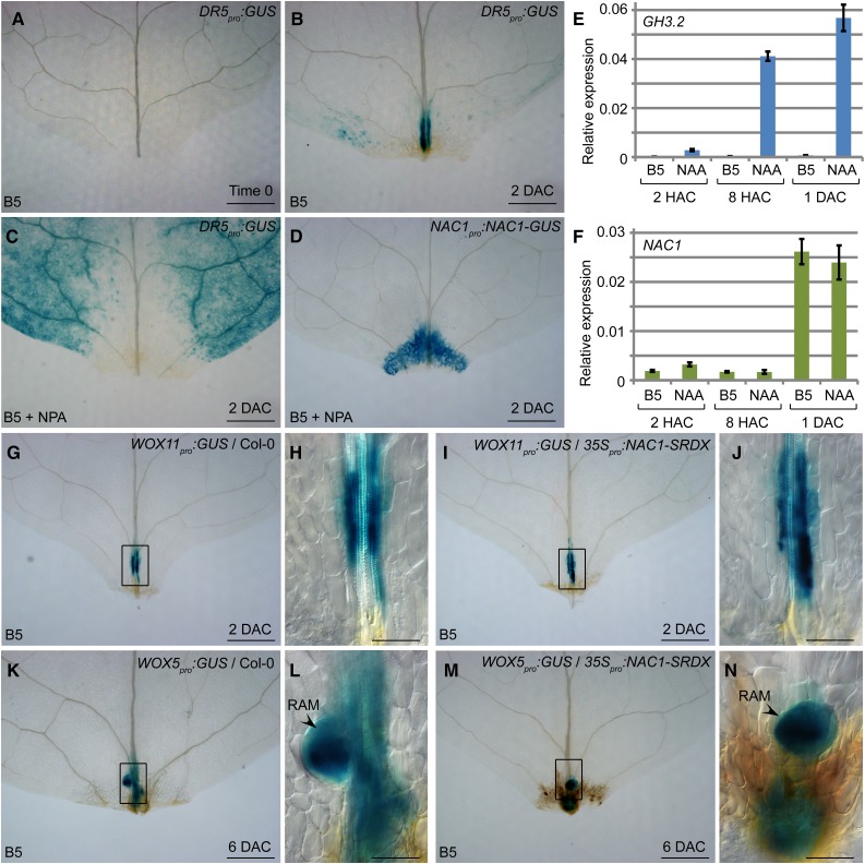
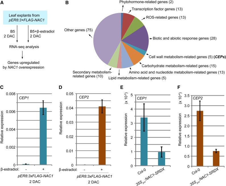
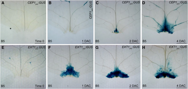
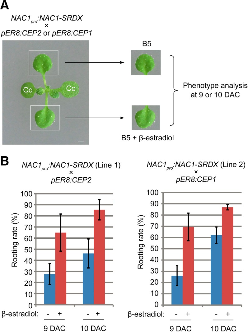
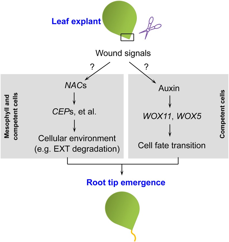
Plants have powerful regenerative abilities that allow them to recover from damage and survive in nature. De novo organogenesis is one type of plant regeneration in which adventitious roots and shoots are produced from wounded and detached organs. By studying de novo root organogenesis using leaf explants of Arabidopsis (Arabidopsis thaliana), we previously suggested that wounding is the first event that provides signals to trigger the whole regenerative process. However, our knowledge of the role of wounding in regeneration remains limited. In this study, we show that wounding not only triggers the auxin-mediated fate transition of regeneration-competent cells, but also induces the NAC pathway for root tip emergence. The NAC1 transcription factor gene was specifically expressed in response to wounding in the leaf explant, but not in the wounded leaf residue of the source plant. Inhibition of the NAC1 pathway severely affected the emergence of adventitious root tips. However, the NAC1 pathway functioned independently of auxin-mediated cell fate transition and regulates expression of CEP genes, which encode proteins that might have a role in degradation of extensin proteins in the cell wall. Overall, our results suggest that wounding has multiple roles in de novo root organogenesis and that NAC1 acts as one downstream branch in regulating the cellular environment for organ emergence.


Figures

Figure 1.
Figure 1.
Identification of NAC genes induced in response to wounding. A, Regeneration system of de novo root organogenesis. B, Plant tissues used in RNA-seq analysis as indicated by the boxed regions. Leaf explant was cut and cultured on B5 medium without carbohydrates in light conditions. Wounded leaf residue was surrounded by air and did not touch the medium. co, cotyledon. C, RNA-seq data showing identification of NAC genes (also see Supplemental Table S1). D and E, qRT-PCR analysis of NAC1 (D) and NAR1 (E) transcript levels in leaf explants and leaf residue compared with those in time-0 leaf as indicated in B. Bars show sd from three PCR experiments. Results were confirmed in three biological repeats. Data from one repeat are shown. Bars = 1 mm in A and B.
Figure 2.
Figure 2.
Expression patterns of NAC genes. A and B, qRT-PCR analysis of NAC1 (A) and NAR1 (B) transcript levels in leaf explants from time 0 to 6 DAC. Bars show sd from three PCR experiments. Results were confirmed in two biological repeats. Data from one repeat are shown. C to E, GUS staining of NAC1pro:NAC1-GUS seedling (C) and 2-DAC leaf explant cultured on B5 medium (D and E). E, Enlargement of wounded region in D. F, GUS staining of NAC1pro:NAC1-GUS leaf explant (upper panel) and wounded leaf residue (lower panel) at 2 DAC. Cutting and culture times as indicated in Fig. 1B. G, Large wound on leaf explant from NAC1pro:NAC1-GUS cultured on B5 medium, showing GUS signal in wounded region. H, Large wound on seedling of NAC1pro:NAC1-GUS, showing no GUS signal in wounded region. I, Stem of NAC1pro:NAC1-GUS was cut, and stem explant (distal part) was cultured on B5 medium while wounded stem residue (proximal part) was still exposed to air. GUS signal was observed only in wounded region of stem explant. J and K, Wounded leaf residue was cultured in B5 medium (J), resulting in NAC1pro:NAC1-GUS signal at wounded region of leaf residue at 4 DAC (K). Bars = 1 mm in C and F to K, 500 μm in D, and 100 μm in E.
Figure 3.
Figure 3.
NAC1 is involved in de novo root organogenesis. A to C, Adventitious rooting from leaf explants of Col-0 (A), NAC1pro:NAC1-SRDX (B), and 35Spro:NAC1-SRDX (C) on B5 medium. Note that both transgenic lines showed defective rooting at 10 DAC. D, Rooting rate analyses of leaf explants from Col-0, NAC1pro:NAC1-SRDX, and 35Spro:NAC1-SRDX. Bars show sd from three biological repeats; n = 30 in each repeat. E, Adventitious rooting from stem explants of Col-0 (left) and 35Spro:NAC1-SRDX (right) cultured on B5 medium, showing rooting defect in 35Spro:NAC1-SRDX. Bars = 1 mm in A to C.
Figure 4.
Figure 4.
NAC1 acts independently of auxin-mediated cell fate transition. A and B, GUS staining of DR5pro:GUS leaf explant cultured on B5 medium at time 0 (A) and 2 DAC (B). C, GUS staining of DR5pro:GUS leaf explant at 2 DAC on medium containing NPA. Note that the GUS signal did not accumulate in wounded region. D, GUS staining of NAC1pro:NAC1-GUS in leaf explant at 2 DAC cultured on B5 medium containing NPA. E and F, qRT-PCR analysis of GH3.2 (E) and NAC1 (F) transcript levels in leaf explants cultured on B5 medium without or with NAA. Bars show sd from three PCR experiments. Results were confirmed in three biological repeats. Data from one repeat are shown. G to J, GUS staining of WOX11pro:GUS (G and H) and WOX11pro:GUS/35Spro:NAC1-SRDX (I and J) leaf explants cultured on B5 medium at 2 DAC. K to N, GUS staining of WOX5pro:GUS (K and L) and WOX5pro:GUS/35Spro:NAC1-SRDX (M and N) leaf explant cultured on B5 medium at 6 DAC. RAM, root apical meristem. H, J, L, and N, Enlargements of boxed regions in G, I, K, and M, respectively. Bars = 500 μm in A to D, G, I, K, and M and 100 μm in H, J, L, and N.
Figure 5.
Figure 5.
NAC1 promotes expression of CEP genes. A, RNA-seq analysis using 2-DAC leaf explants from pER8:3×FLAG-NAC1. Leaf explants were cultured on B5 medium containing β-estradiol/DMSO or DMSO only (control). B, Analysis of upregulated genes in RNA-seq data as indicated in A. Also see Supplemental Table S2. C and D, qRT-PCR analysis of CEP1 (C) and CEP2 (D) transcript levels in leaf explants of pER8:3×FLAG-NAC1 cultured on medium without (−) or with (+) 10 μm β-estradiol at 2 DAC. E and F, qRT-PCR analysis of CEP1 (E) and CEP2 (F) transcript levels in leaf explants of 35Spro:NAC1-SRDX at 4 DAC. Bars in C to F show sd from three PCR experiments. Results were confirmed in two biological repeats. Data from one biological repeat are shown.
Figure 6.
Figure 6.
Expression patterns of CEP1 and EXT1. A to D, GUS staining of CEP1pro:GUS leaf explant cultured on B5 medium at time 0 (A), 1 DAC (B), 2 DAC (C), and 4 DAC (D). E to H, GUS staining of EXT1pro:GUS leaf explant cultured on B5 medium at time 0 (E), 1 DAC (F), 2 DAC (G), and 4 DAC (H). Bars = 500 µm.
Figure 7.
Figure 7.
Overexpression of CEP partially rescues rooting defect in NAC1pro:NAC1-SRDX. A, Design of rescue experiment. Each leaf explant from the same source plant was cultured on B5 medium containing β-estradiol/DMSO or DMSO only (control). co, Cotyledon. B, Rooting rate analyses of leaf explants from two independent crossed lines (NAC1pro:NAC1-SRDX/pER8:CEP2 in the left panel and NAC1pro:NAC1-SRDX/pER8:CEP1 in the right panel) cultured on medium without (−) or with (+) 10 μm β-estradiol at 9 and 10 DAC. Bars show sd with three biological repetitions. n ≥ 22 in each repetition.
Figure 8.
Figure 8.
Model for wounding-induced de novo root organogenesis. Wound signals function in at least two separate pathways for de novo root organogenesis. The molecular features of wound signals remain unclear.

Similar articles

Cited by

References

    1. Aida M, Ishida T, Fukaki H, Fujisawa H, Tasaka M (1997) Genes involved in organ separation in Arabidopsis: an analysis of the cup-shaped cotyledon mutant. Plant Cell 9: 841–857 - PMC - PubMed
    1. Anders S, Huber W (2010) Differential expression analysis for sequence count data. Genome Biol 11: R106. - PMC - PubMed
    1. Anders S, Pyl PT, Huber W (2015) HTSeq--a Python framework to work with high-throughput sequencing data. Bioinformatics 31: 166–169 - PMC - PubMed
    1. Asahina M, Azuma K, Pitaksaringkarn W, Yamazaki T, Mitsuda N, Ohme-Takagi M, Yamaguchi S, Kamiya Y, Okada K, Nishimura T, et al. (2011) Spatially selective hormonal control of RAP2.6L and ANAC071 transcription factors involved in tissue reunion in Arabidopsis. Proc Natl Acad Sci USA 108: 16128–16132 - PMC - PubMed
    1. Bostock RM. (1989) Perspectives on wound healing in resistance to pathogens. Annu Rev Phytopathol 27: 343–371

Publication types

MeSH terms