. 2016 Sep 1;524(13):2577-603.
doi: 10.1002/cne.23978. Epub 2016 Feb 24.

Individual sympathetic postganglionic neurons coinnervate myenteric ganglia and smooth muscle layers in the gastrointestinal tract of the rat

Affiliations

Individual sympathetic postganglionic neurons coinnervate myenteric ganglia and smooth muscle layers in the gastrointestinal tract of the rat

Gary C Walter et al. J Comp Neurol. .

Abstract

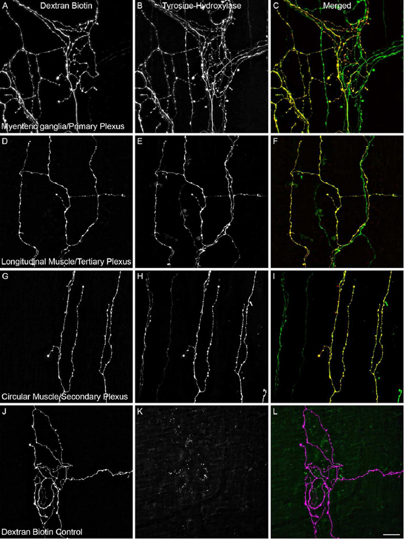
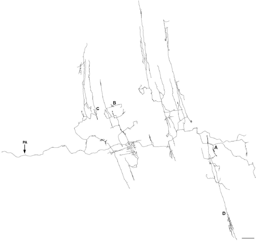
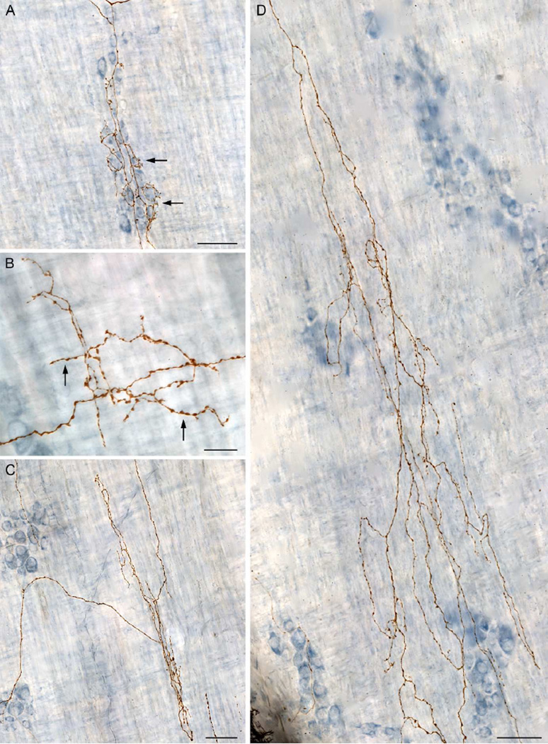
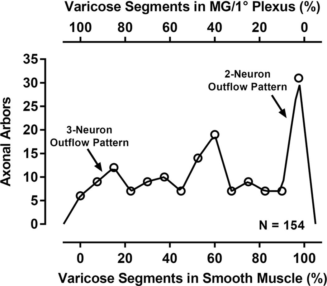
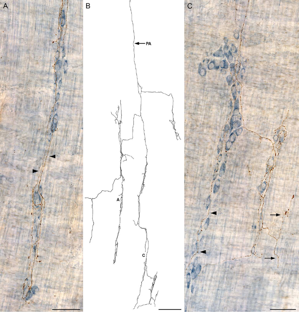
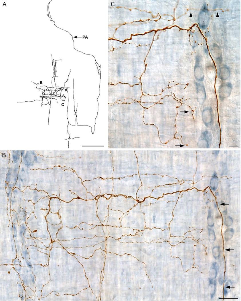
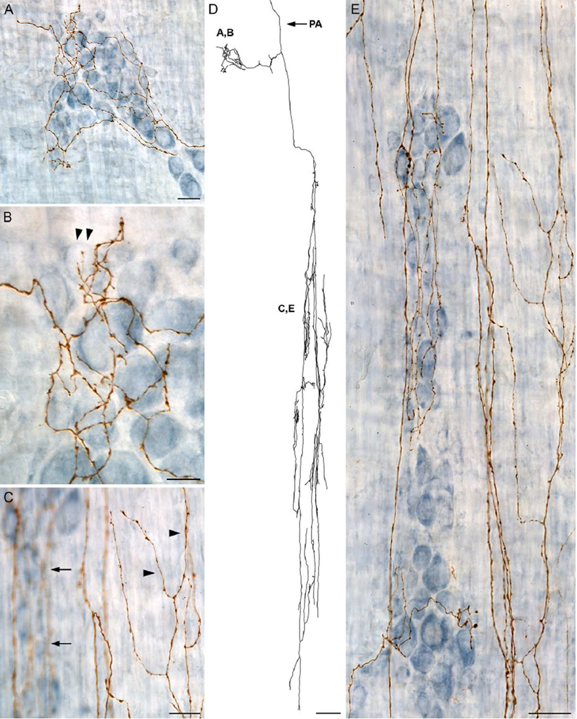
A full description of the terminal architecture of sympathetic axons innervating the gastrointestinal (GI) tract has not been available. To label sympathetic fibers projecting to the gut muscle wall, dextran biotin was injected into the celiac and superior mesenteric ganglia (CSMG) of rats. Nine days postinjection, animals were euthanized and stomachs and small intestines were processed as whole mounts (submucosa and mucosa removed) to examine CSMG efferent terminals. Myenteric neurons were counterstained with Cuprolinic Blue; catecholaminergic axons were stained immunohistochemically for tyrosine hydroxylase. Essentially all dextran-labeled axons (135 of 136 sampled) were tyrosine hydroxylase-positive. Complete postganglionic arbors (n = 154) in the muscle wall were digitized and analyzed morphometrically. Individual sympathetic axons formed complex arbors of varicose neurites within myenteric ganglia/primary plexus and, concomitantly, long rectilinear arrays of neurites within circular muscle/secondary plexus or longitudinal muscle/tertiary plexus. Very few CSMG neurons projected exclusively (i.e., ∼100% of an arbor's varicose branches) to myenteric plexus (∼2%) or smooth muscle (∼14%). With less stringent inclusion criteria (i.e., ≥85% of an axon's varicose branches), larger minorities of neurons projected predominantly to either myenteric plexus (∼13%) or smooth muscle (∼27%). The majority (i.e., ∼60%) of all individual CSMG postganglionics formed mixed, heterotypic arbors that coinnervated extensively (>15% of their varicose branches per target) both myenteric ganglia and smooth muscle. The fact that ∼87% of all sympathetics projected either extensively or even predominantly to smooth muscle, while simultaneously contacting myenteric plexus, is consistent with the view that these neurons control GI muscle directly, if not exclusively. J. Comp. Neurol. 524:2577-2603, 2016. © 2016 Wiley Periodicals, Inc.

Keywords: RRID:AB10566286; RRID:AB_10562715; RRID:AB_2307337; RRID:AB_2313574; RRID:AB_2313713; RRID:AB_2336819; RRID:AB_572268; RRID:RGD_61109; RRID:nig-0000-10294; RRID:rid_000081; autonomic nervous system; celiac ganglion; enteric nervous system; muscularis externa; superior mesenteric ganglion.


Conflict of interest statement

Conflict of Interest Statement

The Authors declare no conflict of interest.

Figures

Figure 1
Figure 1
Labeled arbors located in the gut wall following injection of dextran into the celiac and superior mesenteric ganglia (CSMG) were determined to be predominately catecholaminergic postganglionic neurites by the presence of the noradrenaline synthesizing enzyme tyrosine hydroxylase (TH). Dextran biotin (left column; A,D,G) labeled neurites were co-localized with TH (middle column; B,E,H) as demonstrated in the merged images (right colum; C,F,I; green neurites are dextran-negative/TH-positive while yellow neurites are dextran-positive/TH-positive) throughout the three target tissues surveyed. ALEXA Fluor 594 labeled dextran-positive neurites (J; and purple labeling in panel L) were absent when the tissue was viewed with the QMAX-Green filter in place (K). Spectral bleed-through was similarly ruled out for the ALEXA Fluor 488 fluorophore by the presence of TH-positive/dextran-negative (green) neurites visible in the merged panels (C,F,I). Scale bar in L = 20 µm.
Figure 2
Figure 2
A Neurolucida tracing, collected at 630×, of a single sympathetic neurite with terminal branches and/or varicose fibers of passage located in the myenteric ganglia/1° plexus, the circular muscle/2° plexus, and the longitudinal muscle/3° plexus. The parent axon (PA) is indicated with an arrow. Figure 3 consists of photomicrographs that correspond to the demarcated areas on the tracing. Scale bar = 500 µm.
Figure 3
Figure 3
Images were taken from the traced dextran-biotin labeled ending shown in Figure 2. A: Innervation of the myenteric ganglia/1° plexus by varicose neurites (arrows). B: Varicose fibers terminating in the tertiary plexus (arrows). C: Varicose fibers terminating in the circular muscle/2° plexus. D: Varicose fibers innervating the circular muscle. The dextran-biotin labeled neurites were visualized using DAB (brown) and the myenteric neurons were counterstained with Cuprolinic Blue (blue). Scale bars in A, C, D = 50 µm; B = 20 µm.
Figure 4
Figure 4
The distribution of the complete sample of 154 CSMG postganglionic arbors (number of arbors on y-axis) plotted as a function of the percent of each arbor’s varicose branches that contacted smooth muscle tissue sites (see lower x-axis) or, conversely, that contacted myenteric ganglion/1° plexus tissue sites (see upper x-axis). The majority of sympathetic arbors did not conform to either a strict two-neuron outflow model or a strict three-neuron outflow model. MG/1° Plexus = myenteric ganglia/1° plexus.
Figure 5
Figure 5
A dextran-biotin labeled sympathetic axon that innervated predominately the myenteric ganglia/1° plexus. A: Multiple ganglia within the myenteric plexus were innervated with interconnective strand elements (arrowheads) between the ganglia. B: A Neurolucida tracing of the entire terminal from which panels A and C are taken. The neurite consisted of a long parent axon (PA; entering at 12 o’clock) that repeatedly branched to innervate numerous ganglia and interconnective strands of the myenteric plexus. C: Varicose fibers can be seen within the ganglia and passing through the ganglia (arrowheads). A small spur innervates the circular muscle/2° plexus (arrows). Scale bars in A & C = 63 µm; B = 250 µm.
Figure 6
Figure 6
A dextran-biotin labeled sympathetic axon that innervates the myenteric ganglia/1° plexus, the circular muscle/2° plexus, and the longitudinal muscle/3° plexus. A: The prominent feature of the traced neurite, which is shown in its entirety, was the extensive innervation of the longitudinal muscle/3° plexus between parallel myenteric ganglia (dark blue stained neurons shown in panel B). The parent axon (PA) is indicated with an arrow. B: The smooth large diameter parent axon (arrows) passes through the myenteric ganglia/1° plexus (dark blue neurons), makes a 90 degree turn, and enters the longitudinal muscle/3° plexus where it branches extensively into small caliber varicose axons that run predominately perpendicular to the myenteric ganglia. C: A higher power image of the myenteric ganglia/1° plexus (dark blue neurons), shown in panel B, with the ganglia in focus to illustrate how the small diameter, varicose axons innervating the longitudinal muscle/3° plexus pass both underneath the myenteric ganglia (slightly out of focus axons; arrowheads) and along the border of myenteric ganglia (in focus axons; arrows). Scale bars in A = 250 µm; B = 31 µm; C = 10 µm.
Figure 7
Figure 7
A dextran-biotin labeled sympathetic axon that innervates the myenteric ganglia/1° plexus and the circular muscle/2° plexus. A: Upon innervating a ganglion, a neurite branches extensively giving off numerous collateral varicose processes. B: A high power image of one pole of the ganglia illustrating the close association between varicose processes and the myenteric neurons that they encircle (arrowheads). C: Innervation of the circular muscle/2° plexus is shown in focus (arrowheads) while the innervation of the myenteric plexus by collaterals from the same parent axon are intentionally shown out of focus (arrows) to illustrate the targeting of the different tissues by the same neurite. D: A tracing of the entire ending highlights the morphology of the long rectilinear multi-branching arrays that run within the circular muscle and superficial to the circular muscle within the secondary plexus. The parent axon (PA) is indicated with an arrow. E: An all-in-focus image of an axonal array innervating the circular muscle/2° plexus with collateral axons produced by the same sympathetic neuron innervating the myenteric plexus which runs parallel to the circular muscle. Scale bars in A & C = 20 µm; B = 10 µm; D = 125 µm; E = 31 µm.
Figure 8
Figure 8
A: The number of myenteric ganglia innervated by terminal branches was greatest in the proximal gut and decreased in the distal gut (P = 0.0001). B: The pattern was similar for varicose arbor length (P = 0.0009). Asterisks indicate a significant difference (P < 0.05) compared to the stomach * or duodenum 0–3 cm **; Tukey’s HSD. Mean ± S.E.M.
Figure 9
Figure 9
Sympathetic innervation of the blood vessels was observed in some of the whole mounts. A: Sympathetic axons silhouette the outer perimeter of a blood vessel before giving off several collateral branches that extend into the neighboring myenteric ganglia. B: A tracing of the entire ending documents how the parent axon (PA; arrow) bifurcates and the resulting secondary fibers then run parallel in close association to the blood vessel with several collateral branches innervating the nearby myenteric ganglia. C: At higher magnification, the axons in close apposition to the outer wall of a blood vessel are clearly varicose (arrows). D: The innervation of the myenteric ganglia is similarly varicose (arrows). The brown-stained irregular structures in panels A and C are presumably erythrocytes that were not exsanguinated during perfusion and fixation. Scale bars in A = 125 µm; B = 250 µm; C & D = 20 µm.

Similar articles

Cited by

References

    1. Baker DM, Santer RM. Image analysis of the sympathetic innervation of the myenteric plexus in the small intestine of mammalian species. Comp Biochem Physiol C. 1989;94(2):527–531. - PubMed
    1. Baluk P, Gabella G. Scanning electron microscopy of the muscle coat of the guinea-pig small intestine. Cell Tissue Res. 1987;250(3):551–561. - PubMed
    1. Bar-Shai A, Maayan C, Vromen A, Udassin R, Nissan A, Freund HR, Hanani M. Decreased density of ganglia and neurons in the myenteric plexus of familial dysautonomia patients. J Neurol Sci. 2004;220(1–2):89–94. - PubMed
    1. Bennett MR. Autonomic neuromuscular transmission. viii. Cambridge Eng.: University Press; 1972. p. 273.
    1. Berthoud HR, Patterson LM, Neumann F, Neuhuber WL. Distribution and structure of vagal afferent intraganglionic laminar endings (IGLEs) in the rat gastrointestinal tract. Anat Embryol (Berl) 1997;195(2):183–191. - PubMed

Publication types

MeSH terms