

The association between MR T1ρ and T2 of cartilage and patient-reported outcomes after ACL injury and reconstruction
- PMID: 26850823
- PMCID: PMC4907855
- DOI: 10.1016/j.joca.2016.01.985
The association between MR T1ρ and T2 of cartilage and patient-reported outcomes after ACL injury and reconstruction
Abstract
Objective: To determine if cartilage T1ρ and T2 relaxation time measures after ACL injury and prior to reconstruction (baseline) are associated with patient-reported outcomes at baseline, 6-months, and 1-year after surgery.
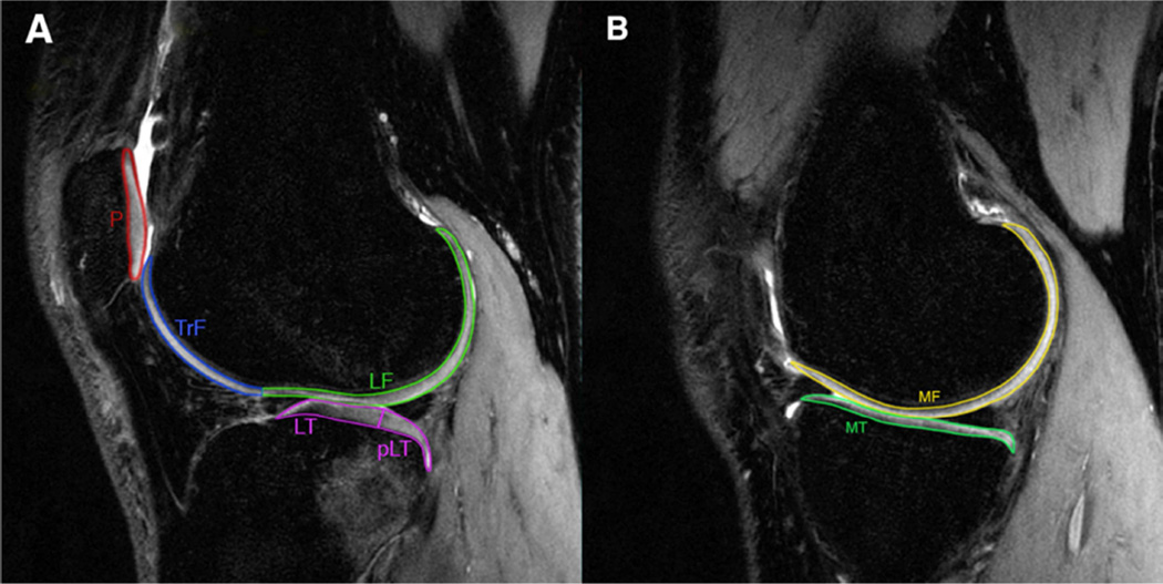
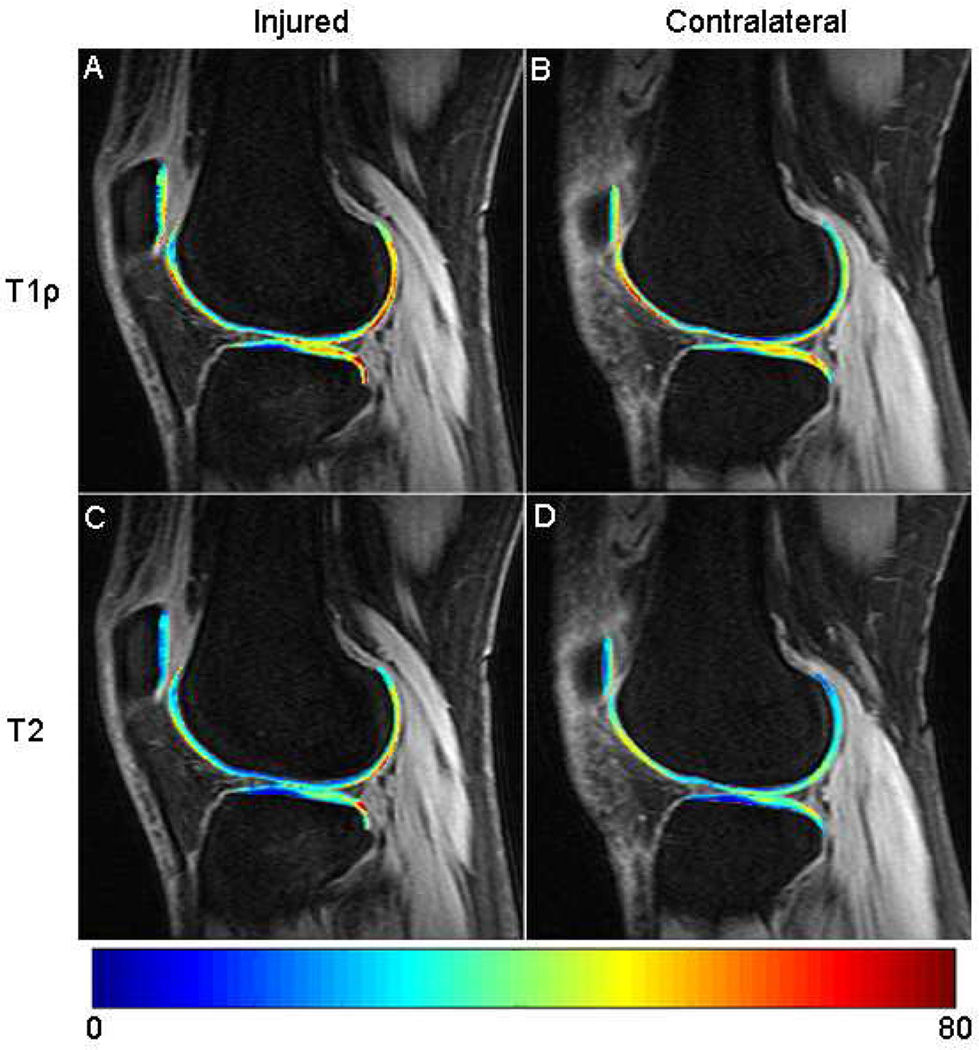
Design: Fifty-four ACL-injured participants were scanned in both knees at baseline using 3T MR T1ρ and T2 mapping. Participants also completed Knee-injury and Osteoarthritis Outcome Score (KOOS) and Marx activity level questionnaires at baseline, 6-months, and 1-year after reconstruction. The difference between cartilage T1ρ or T2 of the injured and contralateral knee (side-to-side difference, SSD) was calculated to account for physiological variations among patients. Linear regression models were built to evaluate the association between the baseline SSD T1ρ or T2 and KOOS or Marx at all time points.
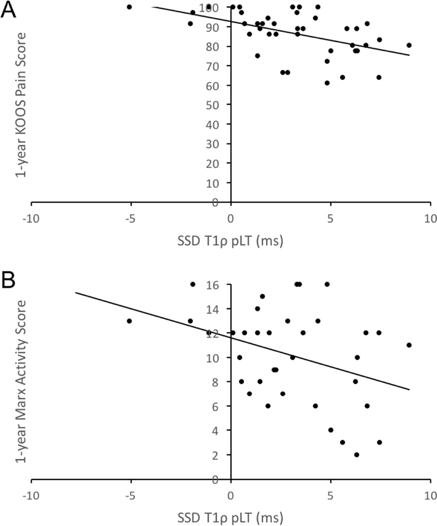
Results: Higher baseline SSD T1ρ posterolateral tibia (pLT) was associated with worse KOOS in all subscales except symptoms at baseline, worse KOOS pain at 6-months, and worse KOOS in all subscales except sports function at 1-year. Higher baseline SSD T2 femoral trochlea (TrF) was associated with worse KOOS activities of daily living (ADL) at 1-year. Higher baseline SSD T1ρ pLT was associated with lower Marx activity level at 1-year. More severe cartilage lesions, as assessed by Whole-Organ MRI Scoring (WORMS), was significantly associated with worse KOOS pain at 6-months and 1-year.
Conclusion: T1ρ and T2 of cartilage after ACL injury were associated with KOOS after injury and both KOOS and Marx after reconstruction. Such associations may help clinicians stratify outcomes post-injury, and thus, improve patient management.
Keywords: ACL reconstruction; Cartilage; KOOS; Marx activity rating scale; T1ρ; T2.
Copyright © 2016 Osteoarthritis Research Society International. Published by Elsevier Ltd. All rights reserved.
Figures
References
-
- Brophy RH, Zeltser D, Wright RW, Flanigan D. Anterior cruciate ligament reconstruction and concomitant articular cartilage injury: incidence and treatment. Arthroscopy. 2010;26:112–120. - PubMed
-
- Sanders TG, Medynski MA, Feller JF, Lawhorn KW. Bone contusion patterns of the knee at MR imaging: footprint of the mechanism of injury. Radiographics. 2000;20 Spec No:S135–S151. - PubMed
-
- Hanypsiak BT, Spindler KP, Rothrock CR, Calabrese GJ, Richmond B, Herrenbruck TM, et al. Twelve-year follow-up on anterior cruciate ligament reconstruction: long-term outcomes of prospectively studied osseous and articular injuries. Am J Sports Med. 2008;36:671–677. - PubMed
-
- Spindler KP, Schils JP, Bergfeld JA, Andrish JT, Weiker GG, Anderson TE, et al. Prospective study of osseous, articular, and meniscal lesions in recent anterior cruciate ligament tears by magnetic resonance imaging and arthroscopy. Am J Sports Med. 1993;21:551–557. - PubMed
-
- Piasecki DP, Spindler KP, Warren TA, Andrish JT, Parker RD. Intraarticular injuries associated with anterior cruciate ligament tear: findings at ligament reconstruction in high school and recreational athletes An analysis of sex-based differences. Am J Sports Med. 2003;31:601–605. - PubMed
MeSH terms
Grants and funding
LinkOut - more resources
Full Text Sources
Other Literature Sources
