

N-Arylacyl O-sulfonated aminoglycosides as novel inhibitors of human neutrophil elastase, cathepsin G and proteinase 3
- PMID: 26850997
- PMCID: PMC4976519
- DOI: 10.1093/glycob/cww011
N-Arylacyl O-sulfonated aminoglycosides as novel inhibitors of human neutrophil elastase, cathepsin G and proteinase 3
Abstract
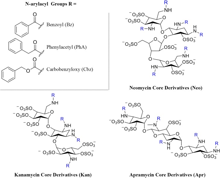
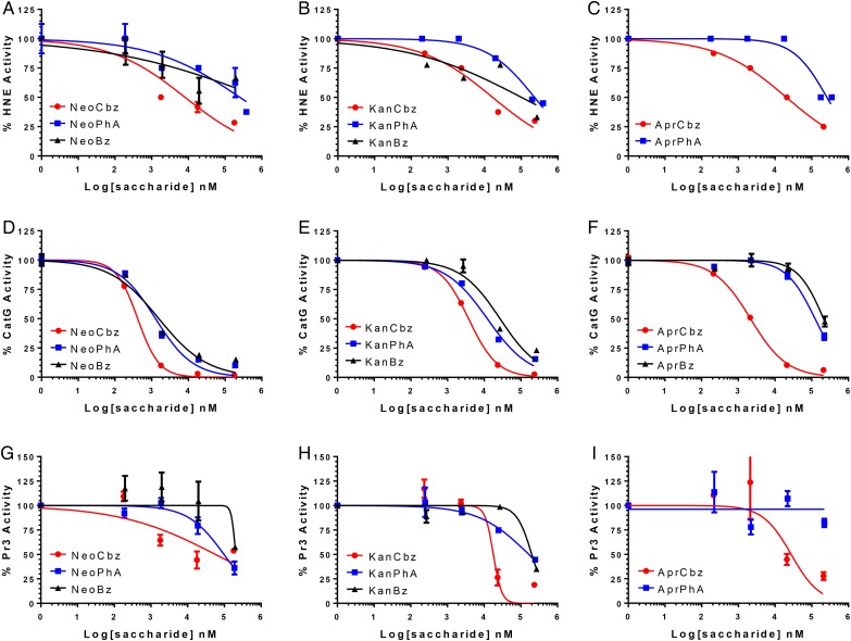
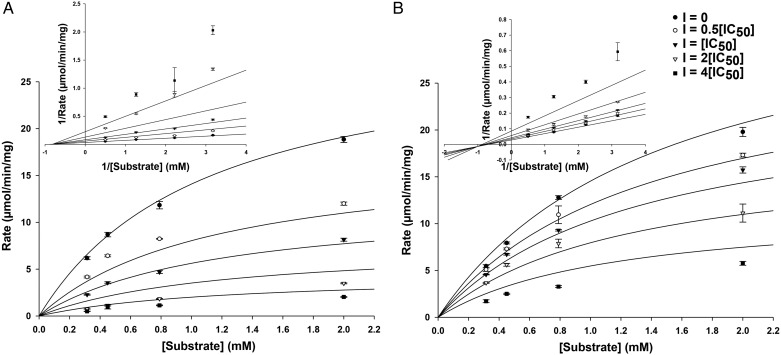
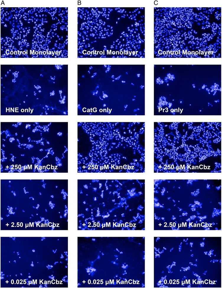
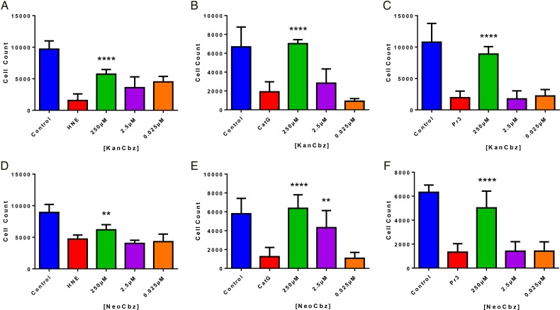
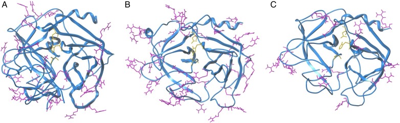
The balance between neutrophil serine proteases (NSPs) and protease inhibitors (PIs) in the lung is a critical determinant for a number of chronic inflammatory lung diseases such as chronic obstructive pulmonary disease, cystic fibrosis and acute lung injury. During activation at inflammatory sites, excessive release of NSPs such as human neutrophil elastase (HNE), proteinase 3 (Pr3) and cathepsin G (CatG), leads to destruction of the lung matrix and continued propagation of acute inflammation. Under normal conditions, PIs counteract these effects by inactivating NSPs; however, in chronic inflammatory lung diseases, there are insufficient amounts of PIs to mitigate damage. Therapeutic strategies are needed to modulate excessive NSP activity for the clinical management of chronic inflammatory lung diseases. In the study reported here, a panel of N-arylacyl O-sulfonated aminoglycosides was screened to identify inhibitors of the NSPs. Dose-dependent inhibitors for each individual serine protease were identified. Select compounds were found to inhibit multiple NSPs, including one lead structure that is shown to inhibit all three NSPs. Two lead compounds identified during the screen for each individual NSP were further characterized as partial mixed inhibitors of CatG. Concentration-dependent inhibition of protease-mediated detachment of lung epithelial cells is demonstrated.
Keywords: glycosaminoglycans; heparin mimics; inflammatory lung disease; neutrophil serine proteases; protease inhibitors.
© The Author 2016. Published by Oxford University Press. All rights reserved. For permissions, please e-mail: journals.permissions@oup.com.
Figures
Similar articles
-
Unopposed cathepsin G, neutrophil elastase, and proteinase 3 cause severe lung damage and emphysema.Am J Pathol. 2014 Aug;184(8):2197-210. doi: 10.1016/j.ajpath.2014.04.015. Epub 2014 Jun 12. Am J Pathol. 2014. PMID: 24929239
-
Using a Caesalpinia echinata Lam. protease inhibitor as a tool for studying the roles of neutrophil elastase, cathepsin G and proteinase 3 in pulmonary edema.Phytochemistry. 2013 Dec;96:235-43. doi: 10.1016/j.phytochem.2013.09.025. Epub 2013 Oct 17. Phytochemistry. 2013. PMID: 24140156
-
Synthesis and pharmacological characterization of 2-aminobenzaldehyde oxime analogs as dual inhibitors of neutrophil elastase and proteinase 3.Bioorg Med Chem. 2015 Mar 1;23(5):1123-34. doi: 10.1016/j.bmc.2014.12.056. Epub 2015 Jan 16. Bioorg Med Chem. 2015. PMID: 25650311
-
SLPI and trappin-2 as therapeutic agents to target airway serine proteases in inflammatory lung diseases: current and future directions.Biochem Soc Trans. 2011 Oct;39(5):1441-6. doi: 10.1042/BST0391441. Biochem Soc Trans. 2011. PMID: 21936830 Review.
-
Neutrophil serine proteinases and defensins in chronic obstructive pulmonary disease: effects on pulmonary epithelium.Eur Respir J. 1998 Nov;12(5):1200-8. doi: 10.1183/09031936.98.12051200. Eur Respir J. 1998. PMID: 9864022 Review.
Cited by
-
Sulphated penta-galloyl glucopyranoside (SPGG) is glycosaminoglycan mimetic allosteric inhibitor of cathepsin G.RPS Pharm Pharmacol Rep. 2023 Jan 6;2(1):rqad001. doi: 10.1093/rpsppr/rqad001. eCollection 2023 Jan. RPS Pharm Pharmacol Rep. 2023. PMID: 36844783 Free PMC article.
-
Integration of Genome-Wide DNA Methylation and Transcription Uncovered Aberrant Methylation-Regulated Genes and Pathways in the Peripheral Blood Mononuclear Cells of Systemic Sclerosis.Int J Rheumatol. 2018 Sep 2;2018:7342472. doi: 10.1155/2018/7342472. eCollection 2018. Int J Rheumatol. 2018. PMID: 30245726 Free PMC article.
-
On the Process of Discovering Leads That Target the Heparin-Binding Site of Neutrophil Elastase in the Sputum of Cystic Fibrosis Patients.J Med Chem. 2019 Jun 13;62(11):5501-5511. doi: 10.1021/acs.jmedchem.9b00379. Epub 2019 May 28. J Med Chem. 2019. PMID: 31074986 Free PMC article.
-
Computational and Preclinical Analysis of 2-(4-Methyl)benzylidene-4,7-dimethyl Indan-1-one (IPX-18): A Novel Arylidene Indanone Small Molecule with Anti-Inflammatory Activity via NF-κB and Nrf2 Signaling.Biomedicines. 2023 Feb 27;11(3):716. doi: 10.3390/biomedicines11030716. Biomedicines. 2023. PMID: 36979695 Free PMC article.
-
Alpha-1 Antitrypsin-A Target for MicroRNA-Based Therapeutic Development for Cystic Fibrosis.Int J Mol Sci. 2020 Jan 28;21(3):836. doi: 10.3390/ijms21030836. Int J Mol Sci. 2020. PMID: 32012925 Free PMC article. Review.
References
Publication types
MeSH terms
Substances
Grants and funding
LinkOut - more resources
Full Text Sources
Other Literature Sources
