. 2016 Mar 27;428(6):1142-1164.
doi: 10.1016/j.jmb.2016.01.027. Epub 2016 Feb 2.

Conserved Intramolecular Interactions Maintain Myosin Interacting-Heads Motifs Explaining Tarantula Muscle Super-Relaxed State Structural Basis

Affiliations

Conserved Intramolecular Interactions Maintain Myosin Interacting-Heads Motifs Explaining Tarantula Muscle Super-Relaxed State Structural Basis

Lorenzo Alamo et al. J Mol Biol. .

Abstract

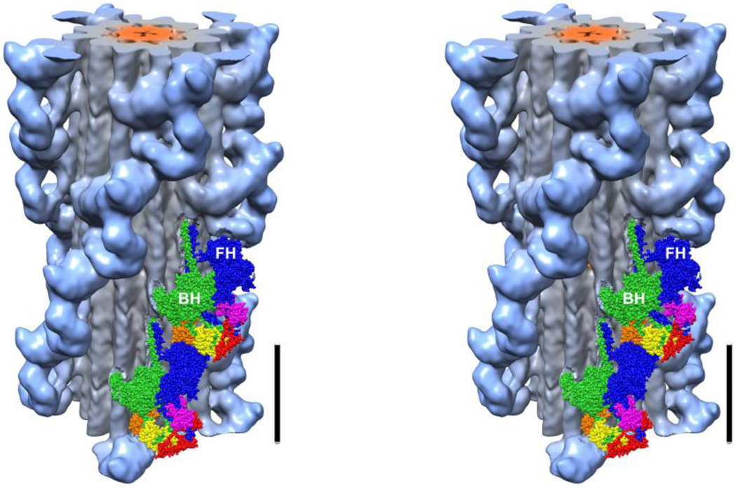
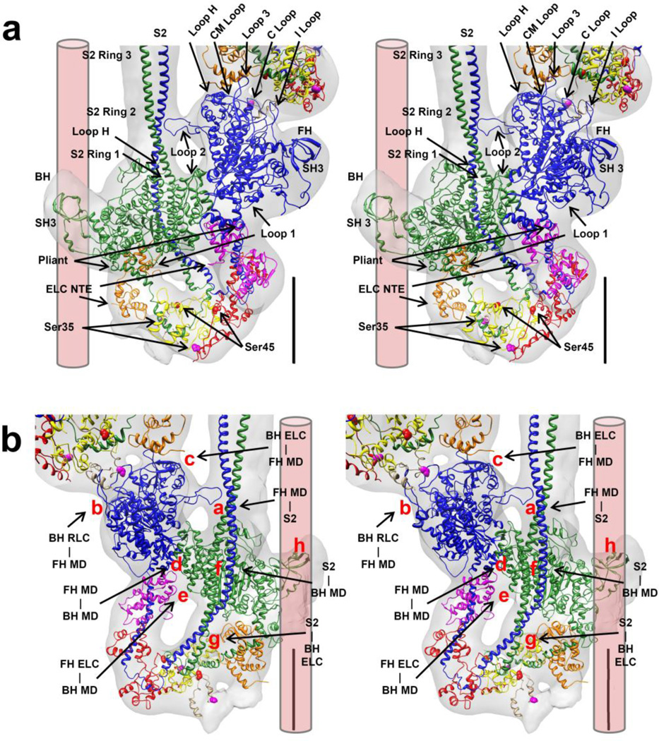
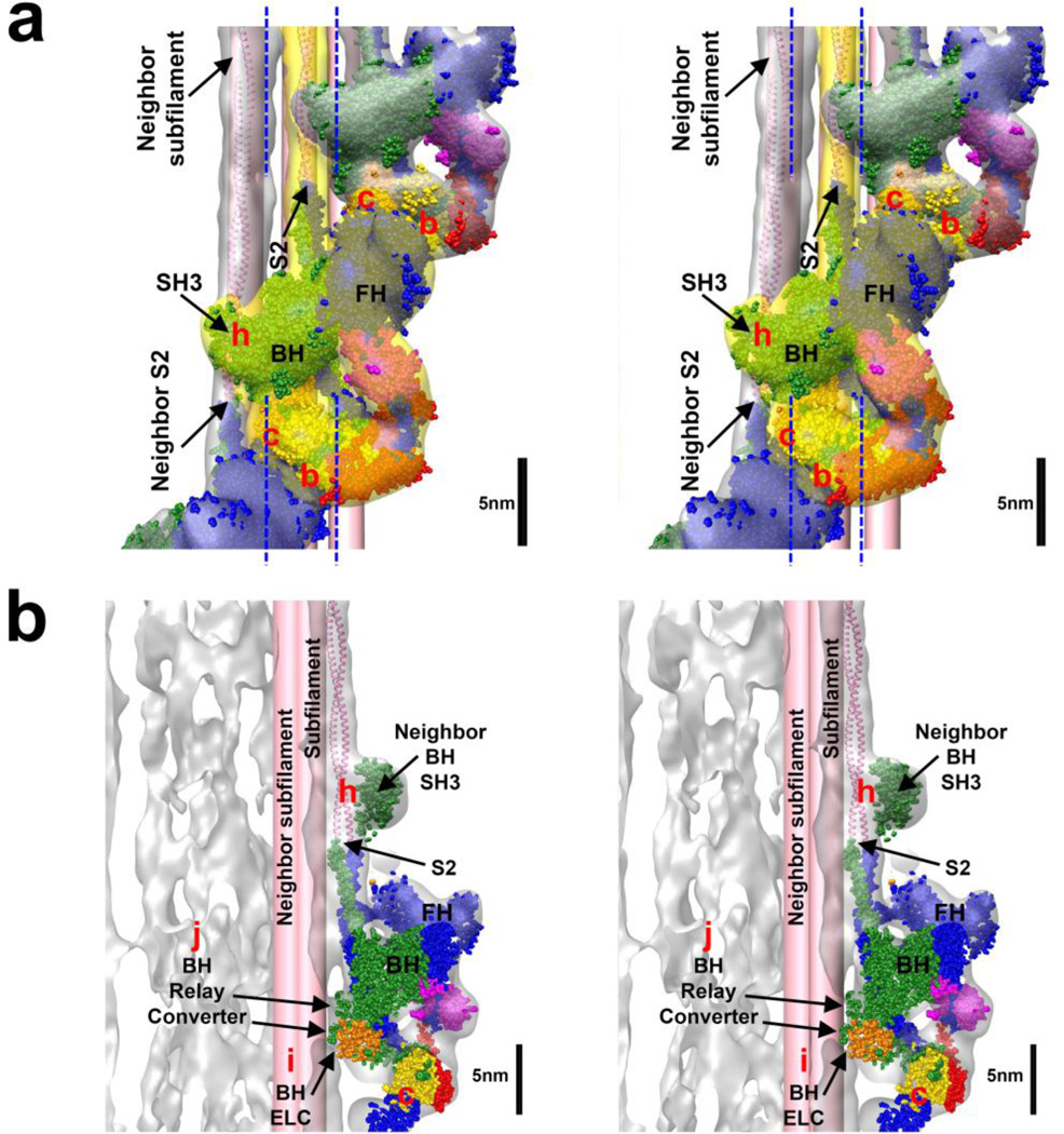
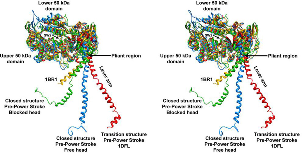
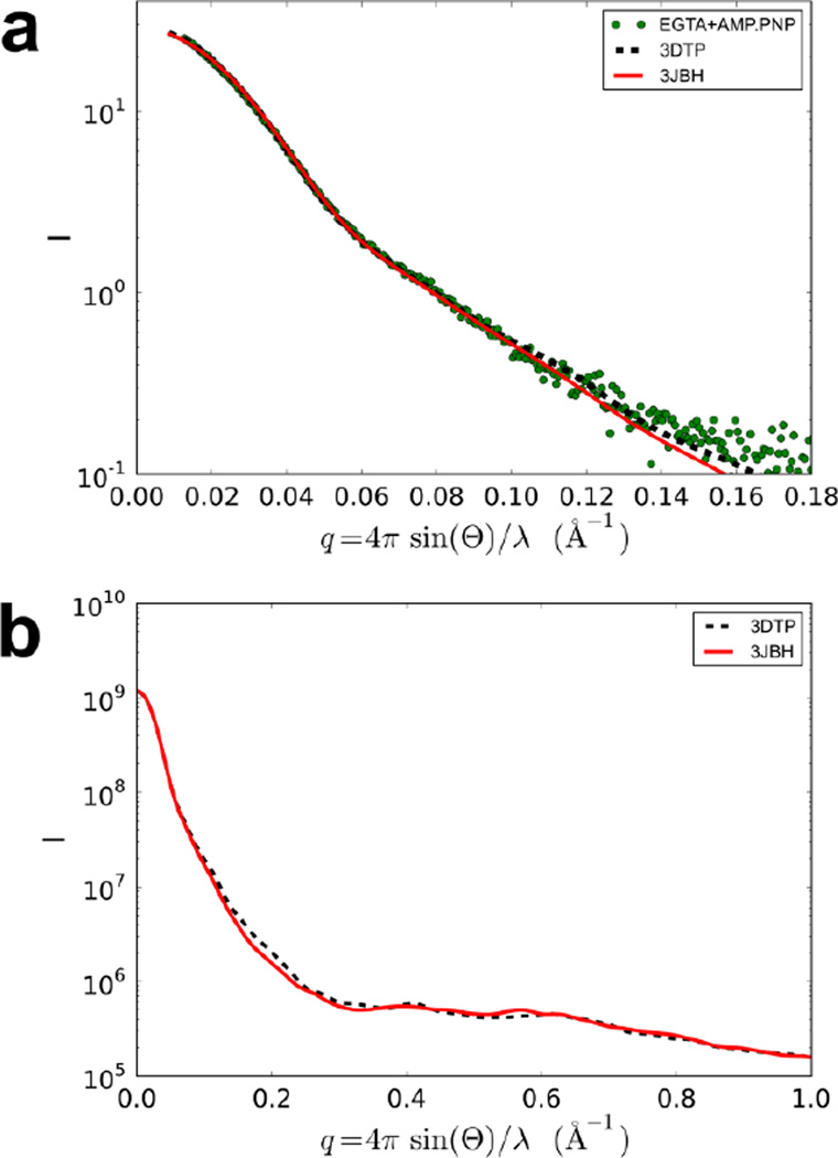
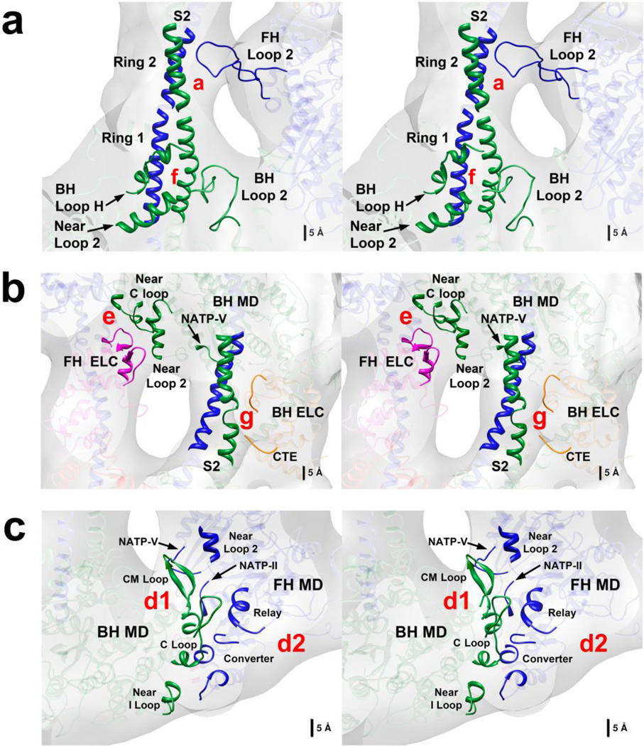
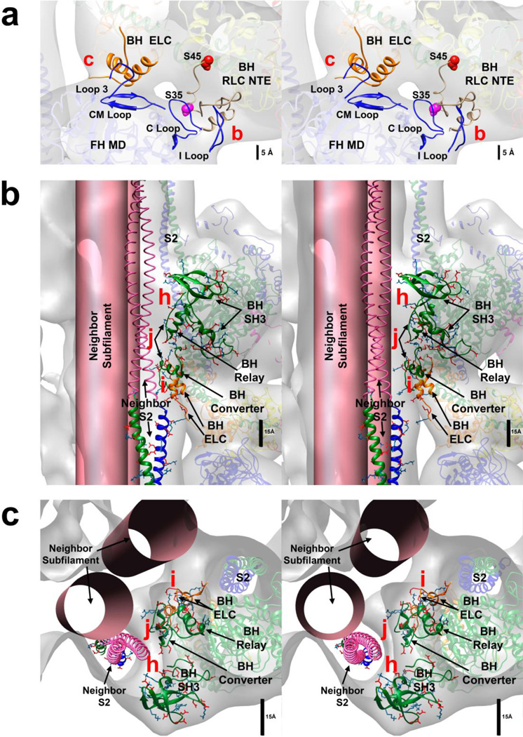
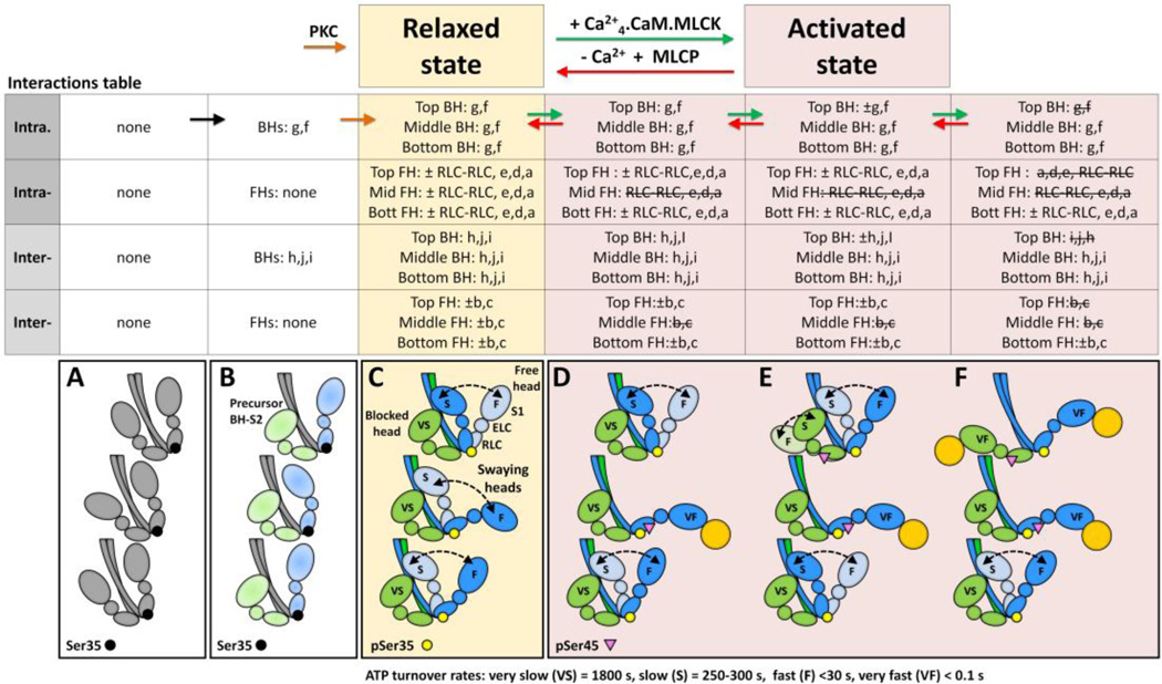
Tarantula striated muscle is an outstanding system for understanding the molecular organization of myosin filaments. Three-dimensional reconstruction based on cryo-electron microscopy images and single-particle image processing revealed that, in a relaxed state, myosin molecules undergo intramolecular head-head interactions, explaining why head activity switches off. The filament model obtained by rigidly docking a chicken smooth muscle myosin structure to the reconstruction was improved by flexibly fitting an atomic model built by mixing structures from different species to a tilt-corrected 2-nm three-dimensional map of frozen-hydrated tarantula thick filament. We used heavy and light chain sequences from tarantula myosin to build a single-species homology model of two heavy meromyosin interacting-heads motifs (IHMs). The flexibly fitted model includes previously missing loops and shows five intramolecular and five intermolecular interactions that keep the IHM in a compact off structure, forming four helical tracks of IHMs around the backbone. The residues involved in these interactions are oppositely charged, and their sequence conservation suggests that IHM is present across animal species. The new model, PDB 3JBH, explains the structural origin of the ATP turnover rates detected in relaxed tarantula muscle by ascribing the very slow rate to docked unphosphorylated heads, the slow rate to phosphorylated docked heads, and the fast rate to phosphorylated undocked heads. The conservation of intramolecular interactions across animal species and the presence of IHM in bilaterians suggest that a super-relaxed state should be maintained, as it plays a role in saving ATP in skeletal, cardiac, and smooth muscles.

Keywords: cryo-electron microscopy; myosin interacting-heads motif; myosin thick filament; striated muscle; super-relaxation.


Figures

Fig. 1
Fig. 1
Wide-eye stereo pair of the longitudinal view of the 3D reconstruction of the frozen-hydrated tarantula thick filament, filtered to 2-nm resolution (EMD-1950) and showing four helical tracks of interacting-heads motifs (IHMs; blue), twelve myosin subfilaments (gray) and the paramyosin core (orange). The 3D map segment shows four 14.5-nm crowns, each of which has four IHMs. The quasi-atomic model PDB 3JBH (formed by two IHMs), which is shown as spheres in the right helix, was flexibly fitted to the 3D map (see Materials and Methods). The myosin heavy chain (MHC) of the blocked head (BH) and free head (FH) are shown in green and blue. The two myosin essential light chains (ELC) are in magenta (FH) or orange (BH). The two myosin regulatory light chains (RLC) are in red (FH) or yellow (BH). Bar: 14.5 nm.
Fig. 2
Fig. 2
Wide-eye stereo pairs of the 2-nm resolution 3D reconstruction of frozen-hydrated relaxed thick filament of tarantula (EMD-1950), shown in grey, with the flexibly fitted quasi-atomic model PDB 3JBH, as viewed from the front (a) or back (b) of the filament surface. The model includes the densities where several loops are located in the blocked head region of interactions “b” and “c” (see Materials and Methods). The MHC in PDB 3JBH shows six surface loops (2, H, CM, 3, C, and I) that are involved in the interactions. Also, the ELC in the PDB 3JBH model shows the extra two amino acids that are missing in the chicken ELC sequence. In (b), the intramolecular interactions are: “a” (FH MD loop 2–S2), “d” (FH MD–BH MD), “e” (FH ELC–BH MD), “f” (S2–BH MD) and “g” (S2–BH ELC). The intermolecular interactions are: “b” (BH RLC–FH MD) and “c” (BH ELC–FH MD), which are established with the adjacent IHM in the filament, and “h,” which occurs between the blocked head SH3 domain and a neighbor myosin S2 (shown as a 2-nm pink cylinder). Each of these interactions is shown in Figs. 6 and 7. MD: motor domain of the myosin head. See legend of Fig. 1. Bar: 50 Å.
Fig. 3
Fig. 3
(a) Wide-eye stereo pairs of three adjacent IHMs forming part of a helix. The intermolecular interactions “b” and “c” are shown with the RLC and ELC of the neighboring blocked head regulatory domain, and interaction “h” is shown with the neighboring S2. The surface of the 3D map corresponding to the IHM in the center is highlighted in yellow. For clarity, the S2 of the model of the two left IHMs has been extended as coiled-coil α-helices (pink). Since the subfilament structure is not known, the two neighboring subfilaments are depicted as cylinders with diameters of about 2.2 nm. (b) Wide-eye stereo pairs of a rotated 90° view of (a), showing that the model of the IHMs is present only in the slice between the two blue dotted lines in (a), causing the neighboring “h” interaction to be far and the “i” and “j” to be closer to the reader. In a relaxed state, the S2 of the IHM emerges from the top with a slight angle of 6°, causing the helix of IHMs to “float,” separated from the backbone surface by about 2 nm. The blocked head is the only part of the IHM that is in contact with the backbone and is covalently connected to it via the S2 and electrostatically connected by three “anchoring” intermolecular interactions: “h” (blocked head SH3 domain) with the extended S2 of an adjacent tail and “i” (blocked head relay/converter) and “j” (blocked head ELC) with the neighboring S2 (see Fig. 7). For a structure color code, see the legend of Fig. 1.
Fig. 4
Fig. 4
Wide-eye stereo pairs of a comparison of the blocked head (green) and free head (blue) of the tarantula IHM PDB 3JBH (Figs. 1–2) with the crystal structures of the pre-power stroke closed PDB 1BR1 (yellow) and transition PDB 1DFL (red). The ELC and RLC were removed to highlight their lever arms, which are in the same plane but have different angles.
Fig. 5
Fig. 5
Small angle X-ray solution scattering (SAXS). Integrated scattering intensity (I in arbitrary units) is given as a function of momentum transfer, q = 4π sin(θ)/λ, with a scattering angle of 2θ and a wavelength of λ. The comparison of model-based (PDB 3DTP and 3JBH) and measured squid HMM scattering profiles in (a) shows that the models cannot be distinguished based on the scattering data that is currently available. The predicted scattering profiles are based on electron microscopy-derived striated tarantula muscle (PDB 3DTP, PDB 3JBH; Fig. 1) IHM models. Calculated wide-angle scattering data (b) confirms that the models do not significantly differ in the wide angle X-ray solution scattering region.
Fig. 6
Fig. 6
Wide-eye stereo pairs showing the general location of intramolecular interactions (a) “a” and “f,” (b) “e” and “g,” and (c) “d,” formed by two sub-interactions, “d.1” and “d.2.” (a) and (c) have the same viewpoint as Fig. 2a and (b) has the viewpoint as Fig. 2b.
Fig. 7
Fig. 7
Wide-eye stereo pairs of (a) intermolecular interactions “c” and “b” and (b) the anchoring intermolecular interactions “h”, “i,” and “j”. For clarity, the neighboring S2 has been extended from the IHM S2 as a coiled-coil α-helix (pink). The neighboring subfilaments, which have unknown structures, are depicted as cylinders with diameters of about 2.2 nm. (c) Stereo pairs of (b) as viewed transversally from the top, showing interactions “h,” “i,” and “j.”
Fig. 8
Fig. 8
Sequential formation, disruption, and reformation of intra- and intermolecular interactions (“interactions table”) in the tarantula IHM PDB 3JBH model upon relaxation (C), activation (C to F, green arrows) and relaxation after activation (F to C, red arrows) according to the cooperative phosphorylation activation (CPA) mechanism (C–F) proposed for tarantula thick filaments allow explanation of the structural basis of the ATP turnover rates detected in tarantula relaxed and Ca2+-activated states. (A) Model of a short segment of a precursor tarantula thick filament showing three precursor IHMs with disordered heads and unphosphorylated Ser35 (black circles). The heads do not make any inter- or intramolecular interactions. (B) Only one head of each precursor IHM in the pre-power stroke closed state (Fig. 4, green) can establish the three anchoring intermolecular interactions and dock them as blocked heads (light green) to the backbone. The free head (light blue), also in the pre-power stroke closed conformation (Fig. 4, blue), can establish intramolecular interactions with the docked blocked head, which are needed to assemble the IHM. (C) In a relaxed state, these precursor IHMs become fully functional after half the Ser35 are monophosphorylated by a temporarily activated protein kinase C (PKC) (brown arrow). PKC can only phosphorylate the fully exposed Ser35 of the free heads (blue heads), as the Ser35 of the blocked heads (green heads) are not accessible., Ser35 monophosphorylation of the free heads allows the free heads to sway away and back by Brownian motion (“swaying” heads) by breaking and reforming the intramolecular interactions (denoted by “±” in the “interactions table” and by dotted curved arrows in the illustration). (C–F) The tarantula CPA mechanism proposed for tarantula thick filament., The interactions table shows how the intra- and intermolecular interactions established in the relaxed state (C) are progressively removed (denoted by crossing them out) upon activation (D–F), ending in a disordered array (F) with fewer interactions. The table also shows how these interactions are progressively reformed from this final disordered array (F) to a newly achieved ordered relaxed array (C). The IHM model and CPA mechanism allow explanation of the structural origin of the very slow (>1800 s), slow (250–300 s) and fast (<30 s) ATP turnover rates detected in tarantula striated muscle in a relaxed state: (1) The very slow rate is associated with unphosphorylated docked blocked heads in the IHM (green heads with motor domains labeled as “VS”) and the few unphosphorylated docked free heads in the IHM (not shown); (2) the slow rate is associated with the Ser35 monophosphorylated free heads (blue heads with motor domains labeled as “S”); and (3) the fast rate is associated with the Ser35 monophosphorylated swaying free heads that are undocked from the IHM (blue heads with motor domains labeled as “F”). Therefore, the docked (helically ordered) unphosphorylated blocked heads and the few free heads with a very slow rate, as well as the transiently docked Ser35 monophosphorylated free heads with a slow rate, are responsible for super-relaxation (SRX) in tarantulas, while the remaining (disordered) swaying free heads that move according to Brownian motion and are undocked from the IHM exhibit the fast rate detected in the disordered relaxed state. In contrast, the very fast (<0.1 s) rate detected in the Ca2+-activated state in tarantula striated muscle is associated with Ser35 monophosphorylated free heads (blue heads with motor domains labeled as “VF”), Ser45 monophosphorylated blocked heads (green heads with motor domains labeled as “VF”), and biphosphorylated free heads (blue heads with motor domains labeled as “VF”) that are bound to actin (yellow spheres) on the activated thin filament. FH: free head, BH: blocked head.

Similar articles

Cited by

References

    1. Craig R, Padrón R. Molecular structure of the sarcomere. In: Engel AG, Franzini-Armstrong C, editors. Myology. 3rd. New York: McGraw-Hill, Inc; 2004. p. 129.p. 166.
    1. Zoghbi ME, Woodhead JL, Moss RL, Craig R. Three-dimensional structure of vertebrate cardiac muscle myosin filaments. Proc. Natl. Acad. Sci. U. S. A. 2008;105:2386–2390. - PMC - PubMed
    1. AL-Khayat HA, Kensler RW, Squire JM, Marston SB, Morris EP. Atomic model of the human cardiac muscle myosin filament. Proc. Natl. Acad. Sci. U. S. A. 2013;110:318–323. - PMC - PubMed
    1. Gonzalez-Sola M, AL-Khayat HA, Behra M, Kensler RW. Zebrafish cardiac muscle thick filaments: isolation technique and three-dimensional structure. Biophys. J. 2014;106:1671–1680. - PMC - PubMed
    1. Woodhead JL, Zhao FQ, Craig R, Egelman EH, Alamo L, Padrón R. Atomic model of a myosin filament in the relaxed state. Nature. 2005;436:1195–1199. - PubMed

Publication types

LinkOut - more resources