. 2016 Apr;1863(4):749-59.
doi: 10.1016/j.bbamcr.2016.02.001. Epub 2016 Feb 2.

Vestiges of Ent3p/Ent5p function in the giardial epsin homolog

Affiliations

Vestiges of Ent3p/Ent5p function in the giardial epsin homolog

Constanza Feliziani et al. Biochim Biophys Acta. 2016 Apr.

Abstract

An accurate way to characterize the functional potential of a protein is to analyze recognized protein domains encoded by the genes in a given group. The epsin N-terminal homology (ENTH) domain is an evolutionarily conserved protein module found primarily in proteins that participate in clathrin-mediated trafficking. In this work, we investigate the function of the single ENTH-containing protein from the protist Giardia lamblia by testing its function in Saccharomyces cerevisiae. This protein, named GlENTHp (for G. lamblia ENTH protein), is involved in Giardia in endocytosis and in protein trafficking from the ER to the vacuoles, fulfilling the function of the ENTH proteins epsin and epsinR, respectively. There are two orthologs of epsin, Ent1p and Ent2p, and two orthologs of epsinR, Ent3p and Ent5p in S. cerevisiae. Although the expression of GlENTHp neither complemented growth in the ent1Δent2Δ mutant nor restored the GFP-Cps1 vacuolar trafficking defect in ent3Δent5Δ, it interfered with the normal function of Ent3/5 in the wild-type strain. The phenotype observed is linked to a defect in Cps1 localization and α-factor mating pheromone maturation. The finding that GlENTHp acts as dominant negative epsinR in yeast cells reinforces the phylogenetic data showing that GlENTHp belongs to the epsinR subfamily present in eukaryotes prior to their evolution into different taxa.

Keywords: ENTH motif; Endocytosis; Giardia lamblia; Vacuole; Vesicle transport; Yeast.


Figures

Figure 1
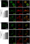
Figure 1. GlENTHp and gENTH expression in S. cerevisiae cells
(A) GlENTHp-mCherry is observed in patches at the plasma membrane in cells expressing the GFP-tagged CLC (upper panels) and wild-type cells (lower panels) incubated with NBD-ceramide. Limited colocalization is observed only between GlENTHp-mCherry and GFP-CLC (inset). Panels on the right show fluorescence and DIC merge. (B) The giardial ENTH domain alone (gENTH) shows cytoplasmic localization. DIC: differential interference contrast. Cells were visualized by confocal microscopy.
Figure 2
Figure 2. GlENTHp and the mutant GlENTHN107Yp are unable to complement ent1Δent2Δ cells
(A) GlENTHp or the gENTH cannot fulfill the essential function of the wild-type yeast epsin. ent1Δent2Δ cells (Δ1Δ2) with an ENT1(TRP1) plasmid and a second URA3 empty plasmid, or encoding the full length GlENTHp, gENTH or ENT1, were grown at 30°C for 3 days on plates containing 5-FAA to evict the ENT1(TRP1) plasmid. (B) Immunoblotting shows GlENTHp-mCherry and gENTH-mCherry, expressed as a ~80 kDa or ~30 kDa band, respectively, in ent1Δent2Δ cells (Δ1Δ2) by using the anti-mCherry Ab. (C) GlENTHN107Yp or its gENTHN107Y mutated domain cannot fulfill the essential function of the wild-type Ent1p. ent1Δent2Δ cells (Δ1Δ2) with an ENT1(TRP1) plasmid and a second URA3 empty plasmid, or encoding the full length GlENTHN107Yp, gENTHN107Y or ENT1p, were grown at 30°C for 3 days on plates containing 5-FAA to evict the ENT1 TRP1 plasmid. (D) Immunoblotting shows GlENTHN107Yp-mCherry and gENTHN107Y-mCherry expressed as a ~80 kDa or ~30 kDa band, respectively, in ent1Δent2Δ cells (Δ1Δ2) by using the anti-Cherry Ab.
Figure 3
Figure 3. GlENTHp cannot act as a dominant negative mutant of epsin in S. cerevisiae cells
(A) Cells expressing GlENTHp show no defect on endocytosis of Ste3-GFP. The wild-type cells expressing Ste3-GFP was transformed with an empty pMET25.426 or with pGlENTHp-mCherry, grown to early log phase on rich medium plates at 30°C and visualized by confocal microscopy. ent1Δent2Δyap1801Δyap1802Δ+Ste3-GFP+ENTH1.414] cells were used as control. The graphic shows the percentage of cells with the observed localization of Ste3. 1: WT+empty vector, 2: WT+GlENTHp-mCh, 3: ent1Δent2Δyap1801Δyap1802Δ (B) Immunoblotting shows GlENTHp-mCherry, expressed as a ~80 kDa band, in Ste3-GFP cells by using the anti-Cherry Ab. (C) Cells expressing GlENTHp show no defect on the actin cytoskeleton. Wild-type cells expressing Abp1-GFP were transformed with an empty pMET25.426 or with pGlENTHp-mCherry(URA3). Abp1-GFP and GlENTHp-mCherry only partially colocalize (insets). [ent1Δent2Δyap1801Δyap1802Δ+ABP1-GFP+ENTH1.414] cells were used as control. Cells were grown to early log phase on rich medium plates at 30°C and visualized by confocal microscopy. DIC: differential interference contrast. Panels on the right show fluorescence and DIC merge. Graphic showing the percentage of cells with the observed Abp1 localization. 1: WT+empty vector, 2: WT+GlENTHp-mCh, 3: ent1Δent2Δyap1801Δyap1802Δ. (D) Immunoblotting shows GlENTHp-mCherry, expressed as a ~80 kDa band, in Abp-GFP cells by using the anti-Cherry Ab.
Figure 4
Figure 4. GlENTHp expression does not restore Golgi-to-vacuole protein trafficking in ent3Δent5Δ cells
(A) GlENTHp cannot compensate the defect in Cps1 intracellular trafficking in ent3Δent5Δ cells. ent3Δent5Δ mutant cells (Δ3Δ5) were co-transformed with a pGFP-Cps1 and the empty vector pMET25.426, pGlENTHp-mCherry or pENT5. ent3Δent5Δ live cells were observed by confocal microscopy. Wild-type cells transformed with pGFP-Cps1 were used as a control (WT). DIC: differential interference contrast. Panels on the right show fluorescence and DIC merge. Graphic showing the percentage of cells with the observed localization of Cps1. 1: WT, 2: Δ3Δ5+empty vector, 3: Δ3Δ5+GlENTHp-mCh, 4: Δ3Δ5+Ent5. (B) GlENTHp cannot restore α-factor maturation. Equivalent OD600 units of wild-type (MATα), wild-type (MATa) (negative control), and ent3Δent5Δ strain (Δ3Δ5) with the empty vector pMET25.426, pGlENTHp-mCherry or pENT5, were spotted on a lawn of supersensitive MATa cells. Halo assay determination of α-factor stimulated signaling and cell cycle arrest activity shows decreased growth inhibition of the underlying lawn for ent3Δent5Δ+GlENTHp-mCherry. (C) Immunoblotting shows GlENTHp-mCherry, expressed as a ~80 kDa band, in ent3Δent5Δ cells by using the anti-Cherry Ab.
Figure 5
Figure 5. GlENTHp functions as an epsinR dominant negative mutant in wild-type S. cerevisiae cells
(A) GlENTHp expression shows defect in Cps1 intracellular trafficking in wild-type cells. Wild-type MATα (WT) cells were co-transformed with a pGFP-Cps1 and with an empty pMET25.423, pGlENTHp-mCherry, pgENTH-mCherry, or pGlENTHK75Ap-mCherry vector. Live cells were observed by confocal microscopy. DIC: differential interference contrast. Panels on the right show fluorescence and DIC merge. Graphic showing the percentage of cells with the observed localization of Cps1. 1: WT+empty vector, 2: WT+GlENTHp-mCh, 3: WT+gENTH-mCh, 4: WT+GlENTHK75Ap-mCh. (B) Only GlENTHp expression reduces α-factor maturation. Equivalent OD600 units of wild-type MATa (WT MATa) cells (negative control) cells with an empty pMET25.426 or pGlENTHp-mCherry and wild-type MATα (WT MATα) (positive control), were spotted on a lawn of supersensitive MATa cells. Halo assay determination shows decreased growth inhibition of the underlying lawn for WT MATα cells expressing GlENTHp-mCherry (top panel) but not when gENTH-mCherry or GlENTHK75Ap-mCherry were expressed (bottom panel). (C) Immunoblotting shows GlENTHp-mCherry (~80 kDa), gENTH-mCherry (~30 kDa) and GlENTHK75Ap-mCherry (~80 kDa) expressed in wild-type (WT/GFP-Cps1) cells by using the anti-Cherry Ab.
Figure 6
Figure 6. The whole GlENTHp produces Ent3/5p misslocalization
(A) Wild-type cells expressing Ent3p-GFP were transformed with pGlENTHp-mCherry, gENTH-mCherry, or pGlENTHK75Ap-mCherry vector. The expression of GlENTHp but not of gENTH or GlENTHK75Ap inhibits the correct localization of Ent3p. Graphic shows the percentage of cells with the observed localization of Ent3p. wt: wild-type cell expressing Ent3p. 1: GlENTHp-mCh, 3: gENTH-mCh, 4: GlENTHK75Ap-mCh. Error bars, s.e.m.; P < 0:005. (B) Wild-type cells expressing Ent5p-GFP were transformed with pGlENTHp-mCherry, gENTH-mCherry, or pGlENTHK75Ap-mCherry vector. Only the expression of GlENTHp causes a localization defect of Ent4p. Live cells were observed by confocal microscopy. Graphic showing the percentage of cells with the observed localization of Ent5p wt: wild-type cell expressing Ent3p or Ent5p. 1: GlENTHp-mCh, 3: gENTH-mCh, 4: GlENTHK75Ap-mCh. Error bars, s.e.m.; P < 0:005.
Figure 7
Figure 7. GlENTHp interacts with adaptor proteins
Strains expressing CLC, APL4, GGA1 and GGA2 transformed with GlENTHp-mCherry were subject to FRET analysis. Representative fluorescence of GFP pre- and postphotobleaching for each strain is showed. Pseudocolor images illustrating FRET efficiency is shown in the right panels. Pseudocolor scale bar is displayed. Bar graphs show the mean FRET efficiency for each pair. Data are presented as mean ± SEM. The number of cells for each group is indicated in parentheses. The pairs GlENTHp/APL4, GlENTHp/GGA1 and GlENTHp/GGA2 showed significant positive FRET when comparing with the negative control of CLC/empty mCherry vector.
Figure 8
Figure 8. Hypothetical model showing how GlENTHp might interfere with the Ent3/5p function
(A) Membrane association of Ent3/5p requires binding of GGAs to PI4P localized in the trans cisternae of the Golgi apparatus (in blue) in yeast and the interaction with AP-1 and clathrin to participate in vesicular Golgi-to-vacuole trafficking (wild-type cells). (B) The whole GlENTH protein competes for PI4P, AP-1 and GGAs binding producing misslocalization of Ent3/5p. However, because GlENTHp do not bind to clathrin, impairs CCV formation and thus unable to complement the Ent3/5p function (wild-type cells overexpressing GlENTHp).

Similar articles

References

    1. Traub LM. Sorting it out: AP-2 and alternate clathrin adaptors in endocytic cargo selection. J Cell Biol. 2003;163:203–208. - PMC - PubMed
    1. Traub LM. Common principles in clathrin-mediated sorting at the Golgi and the plasma membrane. Biochim Biophys Acta. 2005;1744:415–437. - PubMed
    1. Owen DJ, Collins BM, Evans PR. Adaptors for clathrin coats: structure and function. Annu Rev Cell Dev Biol. 2004;20:153–191. - PubMed
    1. Hirst J, Borner GH, Edgar J, Hein MY, Mann M, Buchholz F, Antrobus R, Robinson MS. Interaction between AP-5 and the hereditary spastic paraplegia proteins SPG11 and SPG15. Mol Biol Cell. 2013;24:2558–2569. - PMC - PubMed
    1. Itoh T, De Camilli P. BAR, F-BAR (EFC) and ENTH/ANTH domains in the regulation of membrane-cytosol interfaces and membrane curvature. Biochim Biophys Acta. 2006;1761:897–912. - PubMed

Publication types

MeSH terms

Substances

LinkOut - more resources