. 2017 Jul;66(7):1297-1305.
doi: 10.1136/gutjnl-2015-310409. Epub 2016 Feb 5.

Implantation of healthy matrix-embedded endothelial cells rescues dysfunctional endothelium and ischaemic tissue in liver engraftment

Affiliations

Implantation of healthy matrix-embedded endothelial cells rescues dysfunctional endothelium and ischaemic tissue in liver engraftment

Pedro Melgar-Lesmes et al. Gut. 2017 Jul.

Abstract

Objective: Liver transplantation is limited by ischaemic injury which promotes endothelial cell and hepatocyte dysfunction and eventually organ failure. We sought to understand how endothelial state determines liver recovery after hepatectomy and engraftment.

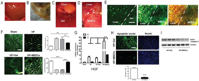
Design: Matrix-embedded endothelial cells (MEECs) with retained healthy phenotype or control acellular matrices were implanted in direct contact with the remaining median lobe of donor mice undergoing partial hepatectomy (70%), or in the interface between the remaining median lobe and an autograft or isograft from the left lobe in hepatectomised recipient mice. Hepatic vascular architecture, DNA fragmentation and apoptosis in the median lobe and grafts, serum markers of liver damage and phenotype of macrophage and lymphocyte subsets in the liver after engraftment were analysed 7 days post-op.

Results: Healthy MEECs create a functional vascular splice in donor and recipient liver after 70% hepatectomy in mouse protecting these livers from ischaemic injury, hepatic congestion and inflammation. Macrophages recruited adjacent to the vascular nodes into the implants switched to an anti-inflammatory and regenerative profile M2. MEECs improved liver function and the rate of liver regeneration and prevented apoptosis in donor liver lobes, autologous grafts and syngeneic engraftment.

Conclusions: Implants with healthy endothelial cells rescue liver donor and recipient endothelium and parenchyma from ischaemic injury after major hepatectomy and engraftment. This study highlights endothelial-hepatocyte crosstalk in hepatic repair and provides a promising new approach to improve regenerative medicine outcomes and liver transplantation.

Keywords: ENDOTHELIAL CELLS; ISCHAEMIA; LIVER IMMUNOLOGY; LIVER REGENERATION.


Conflict of interest statement

Competing interests

The authors declare no competing financial interests.

Figures

Figure 1
Figure 1
Beneficial effects of MEECs preventing liver damage in ischemic median lobe after 70% hepatectomy. C57BL/6 mice underwent 70% hepatectomy (excision of left lobe and half of median lobe). (A) Macroscopic aspect of a pre-op median lobe, 7 days post-op (B) or 7 days post-op with acellular denatured collagen implants (Gel) (C) or (D) MEECs. (E) Vascularity was analyzed in whole liver by angiography (intracardiac perfusion of FITC-dextran, MW 2×10⁶ Da) using intravital multiphoton microscopy. Macrophages were also stained by intravenous injection of 70 kDa Texas red-dextran 2 hours before angiography and sacrifice. Representative images of the vascular network at the interface between the remaining median lobe and denatured collagen or MEECs are shown in green; macrophages are shown in red and intravascular merge of angiography and Texas red-dextran is shown in yellow. (F) Representative images of angiography and quantitative analysis of vascular diameter (congestion) and functional number of vessel branches in the hepatic median lobe of sham or hepatectomized mice (HP) in the presence or absence of acellular implants (HP+Gel) or MEECs (HP+MEECs) 7 days post-op. (G) Gene expression of hepatocyte growth factor (HGF) in ischemic median lobe after 3 or 7 days post-op assessed by Real-time PCR (H) To detect cell death, the terminal deoxynucleotidyl transferase dUTP nick-end labeling (TUNEL) assay was used in median liver lobes from hepatectomized mice in contact with acellular implants or MEECs. Representative images of apoptotic nuclei are shown in green. Nuclei were stained with DAPI in blue. 200x magnification. Quantification of cell death is shown below. (I), Western blot corresponding to active caspase 3 was performed to assess apoptosis in median liver lobes from hepatectomized mice in contact with acellular implants or MEECs. Representative images of three samples of each group to detect active caspase 3 and the housekeeping β–actin are plotted. Scale bars, 100 μm. Data are represented as mean ± s.e.m. **P < 0.01, ***P < 0.001, analysis of variance (ANOVA) or t-student when appropriate.
Figure 2
Figure 2
Vascular and immunomodulatory effects of MEECs in contact with ischemic median lobe improving liver regeneration and function. C57BL/6 mice underwent 70% hepatectomy (excision of left lobe and half of median lobe). (A) Representative images of angiography and quantitative analysis of vascular diameter (congestion) and angiogenesis (number of anastomoses) in the hepatic right lobe of sham or hepatectomized mice (HP) in the presence or absence of acellular implants (HP+Gel) or MEECs (HP+MEECs) 7 days post-op (B) Representative images and quantitative analysis of total number of macrophages and contacts with vessels in the hepatic right lobe analyzed by injection of 70 kDa Texas red-dextran 2 hours before sacrifice and angiography (intracardiac perfusion of FITC-dextran, MW 2×10⁶ Da) using intravital multiphoton microscopy. Macrophages are shown in red and intravascular merge of angiography and Texas red-dextran is shown in yellow. (C) Liver restoration rate was assessed in sham or hepatectomized mice in the presence or absence of acellular implants or MEECs. Liver restoration rate was calculated as liver weight/body weight × 100. (D) Serum markers of liver damage Alanine Aminotransferase (ALT) and Aspartate Aminotransferase (AST) were quantified in hepatectomized mice in the presence of acellular implants or MEECs. Scale bars, 100 μm. Data are represented as mean ± s.e.m. *P < 0.05, **P < 0.01, ***P < 0.001, analysis of variance (ANOVA) or t-student when appropriate.
Figure 3
Figure 3
Hepatic immunomodulation of gene expression profiles of macrophages and T helper lymphocytes 3 or 7 days after implantation of MEECs. Quantification of M1 (iNOS, COX-2 and IL1-β) (A) and M2 (Arg1, MRC1 and Retn1a) (B) gene expression profiles by Real-time PCR in sham or hepatectomized mice in the presence or absence of acellular implants (Gel) or MEECs. (C) Quantification of gene expression profiles of Th1 (INFγ and IL-2) and (D) Th2 (IL-4 and IL-10) by Real-time PCR in sham or hepatectomized mice in the presence or absence of acellular implants (Gel) or MEECs. Data are represented as mean of fold change ± s.e.m. *P < 0.05, **P < 0.01, ***P < 0.001, analysis of variance (ANOVA).
Figure 4
Figure 4
Beneficial effects of MEECs preventing liver damage after autologous engraftment. (A) Schematic representation of surgical implantation of MEECs or acellular implants in the interface between the ischemic median liver lobe and the donated graft from the left liver lobe. (B) Macroscopic aspect of median lobe and autologous grafts implanted with acellular denatured collagen or (C) MEECs 7 days post-op. (D) Vascularity was analyzed in the interface between median liver lobe and autologous graft by angiography using intravital multiphoton microscopy. Representative images of the vascular network at the interface between the remaining median lobe, acellular Denatured collagen or MEECs and the graft are shown in green. (E) Representative images of angiography and quantitative analysis of vascular diameter (congestion) and functional number of vessel branches in the hepatic median lobe of hepatectomized mice in the presence of acellular implants (HP+Gel) or MEECs (HP+MEECs) 7 days post-op. (F) To detect intragraft cell death, TUNEL assay was performed in autologous liver grafts in contact with acellular implants or MEECs. Representative images of apoptotic nuclei are shown in green. Nuclei were stained with DAPI in blue. 200x magnification. Quantification of cell death is shown below. (G) To assess apoptosis, Western blot corresponding to active caspase 3 was performed in autologous liver grafts from mice in contact with acellular implants or MEECs. Representative images of three samples of each group to detect active caspase 3 and the housekeeping β–actin are plotted. (H) Serum markers of liver damage Alanine Aminotransferase (ALT) and Aspartate Aminotransferase (AST) were quantified in hepatectomized mice in the presence of acellular implants or MEECs. Scale bars, 100 μm. Data are represented as mean ± s.e.m. *P < 0.05, **P < 0.01, ***P < 0.001, analysis of variance (ANOVA) or t-student when appropriate.
Figure 5
Figure 5
Beneficial effects of MEECs preventing liver damage after allogeneic engraftment. (A) Macroscopic aspect of median lobe and allogeneic grafts implanted with acellular denatured collagen (Gel) or (B) MEECs 7 days post-op. (C) Vascularity was analyzed in the interface between median liver lobe and allogeneic graft by angiography using intravital multiphoton microscopy. Representative images of the vascular network at the interface between the remaining median lobe, acellular Denatured collagen or MEECs and the graft are shown in green. (D) Representative images of angiography and quantitative analysis of vascular diameter (congestion) and functional number of vessel branches in the hepatic median lobe of hepatectomized mice in the presence of acellular implants (HP+Gel) or MEECs (HP+MEECs) 7 days post-op. (E) To detect intragraft cell death, TUNEL assay was performed in allogeneic liver grafts in contact with acellular implants or MEECs. Representative images of apoptotic nuclei are shown in green. Nuclei were stained with DAPI in blue. 200x magnification. Quantification of cell death is shown below. (F) To assess apoptosis, Western blot corresponding to active caspase 3 was performed in allogeneic liver grafts from mice in contact with acellular implants or MEECs. Representative images of three samples of each group to detect active caspase 3 and the housekeeping β–actin are plotted. (G) Intragraft gene expression profile of immunotolerance expressed as Th1 (INFγ and IL-2) and Th2 (IL-4 and IL-10) cytokine expression analyzed by Real-Time PCR (H) Serum markers of liver damage Alanine Aminotransferase (ALT) and Aspartate Aminotransferase (AST) were quantified in mice with isografts in the presence of acellular implants or MEECs. Scale bars, 100 μm. Data are represented as mean ± s.e.m. *P < 0.05, **P < 0.01, ***P < 0.001, analysis of variance (ANOVA) or t-student when appropriate.

References

    1. Lim YS, Kim WR. The global impact of hepatic fibrosis and end-stage liver disease. Clin Liver Dis. 2008;12:733–746. - PubMed
    1. Zhai Y, Petrowsky H, Hong JC, et al. Ischaemia-reperfusion injury in liver transplantation--from bench to bedside. Nat Rev Gastroenterol Hepatol. 2013;10:79–89. - PMC - PubMed
    1. US Department of Health and Human Services. Organ Procurement and Transplantation Network. 2014 [online], http://optn.transplant.hrsa.gov/data/
    1. Takebe T, Sekine K, Enomura M, et al. Vascularized and functional human liver from an iPSC-derived organ bud transplant. Nature. 2013;499:481–484. - PubMed
    1. Si-Tayeb K, Noto FK, Nagaoka M, et al. Highly efficient generation of human hepatocyte-like cells from induced pluripotent stem cells. Hepatology. 2010;51:297–305. - PMC - PubMed

MeSH terms