

STAT3/5-Dependent IL9 Overexpression Contributes to Neoplastic Cell Survival in Mycosis Fungoides
- PMID: 26851186
- PMCID: PMC4967550
- DOI: 10.1158/1078-0432.CCR-15-1784
STAT3/5-Dependent IL9 Overexpression Contributes to Neoplastic Cell Survival in Mycosis Fungoides
Abstract
Purpose: Sustained inflammation is a key feature of mycosis fungoides (MF), the most common form of cutaneous T-cell lymphoma (CTCL). Resident IL9-producing T cells have been found in skin infections and certain inflammatory skin diseases, but their role in MF is currently unknown.
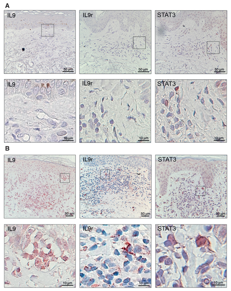
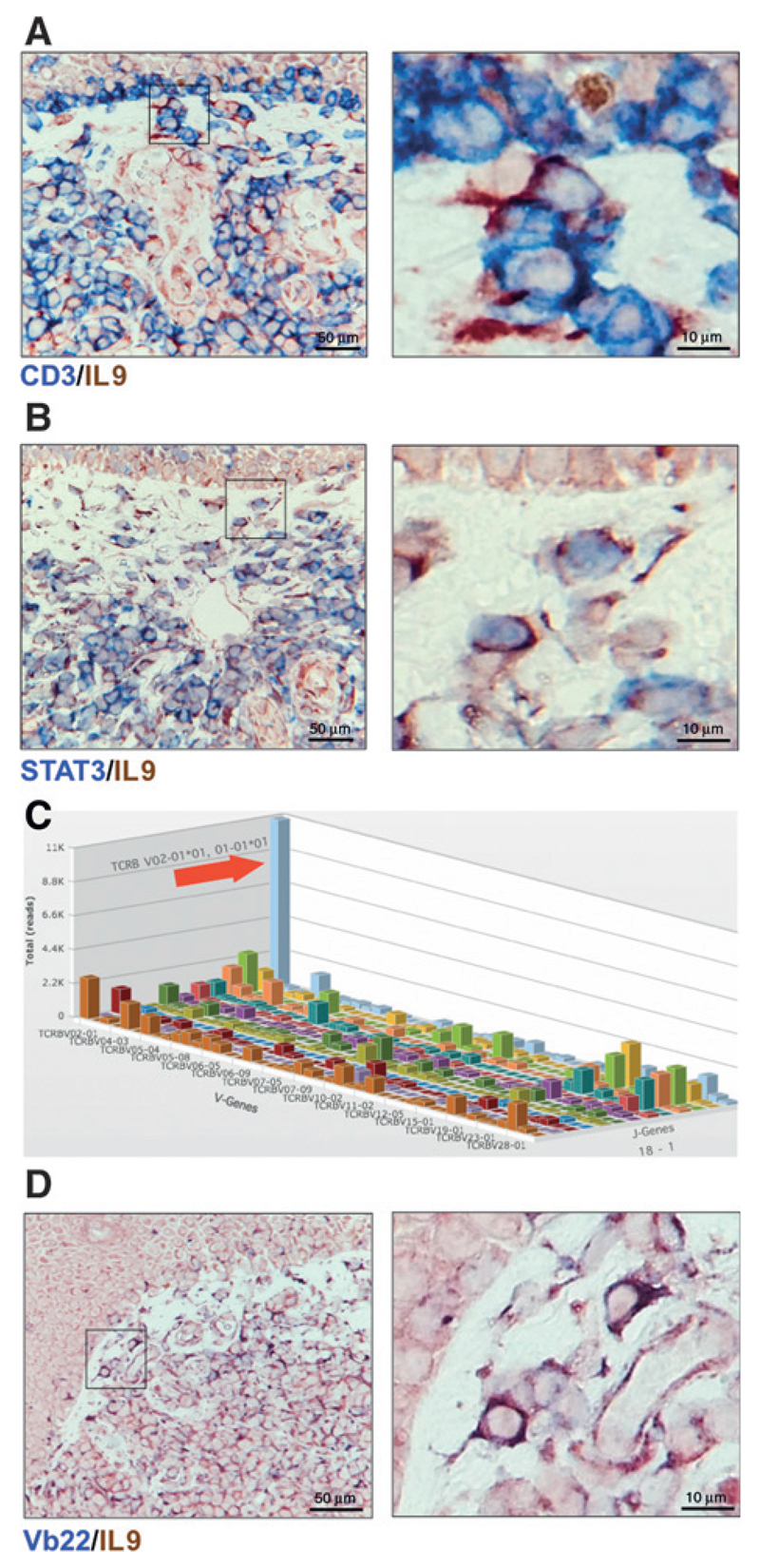
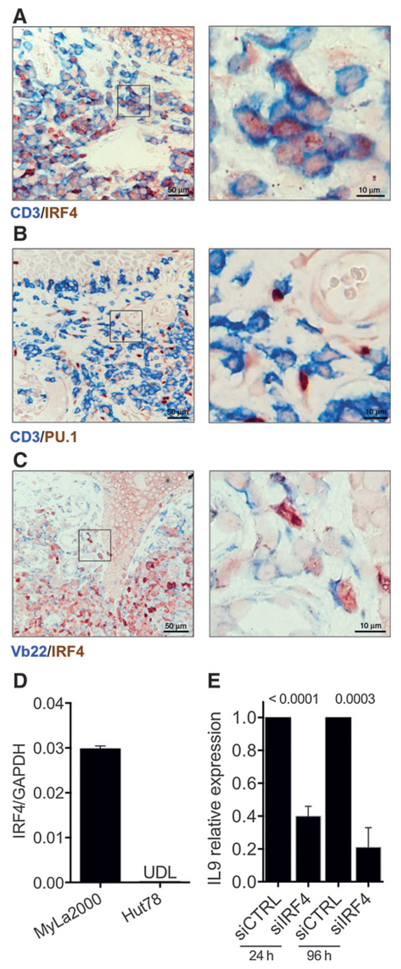
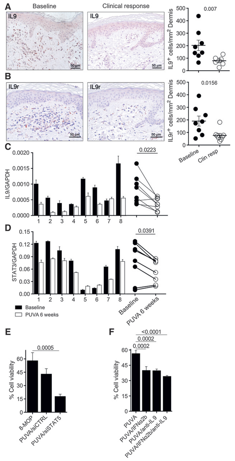
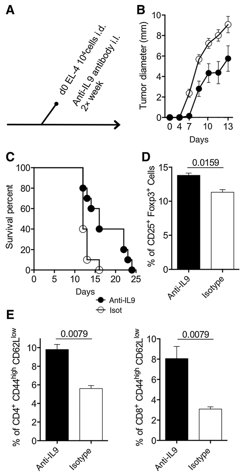
Experimental design: We analyzed lesional skin from patients with MF for the expression of IL9 and its regulators. To determine which cells were producing IL9, high-throughput sequencing was used to identify malignant clones and Vb-specific antibodies were employed to visualize malignant cells in histologic preparations. To explore the mechanism of IL9 secretion, we knocked down STAT3/5 and IRF4 by siRNA transfection in CTCL cell lines receiving psoralen+UVA (PUVA) ± anti-IL9 antibody. To further examine the role of IL9 in tumor development, the EL-4 T-cell lymphoma model was used in C57BL/6 mice.
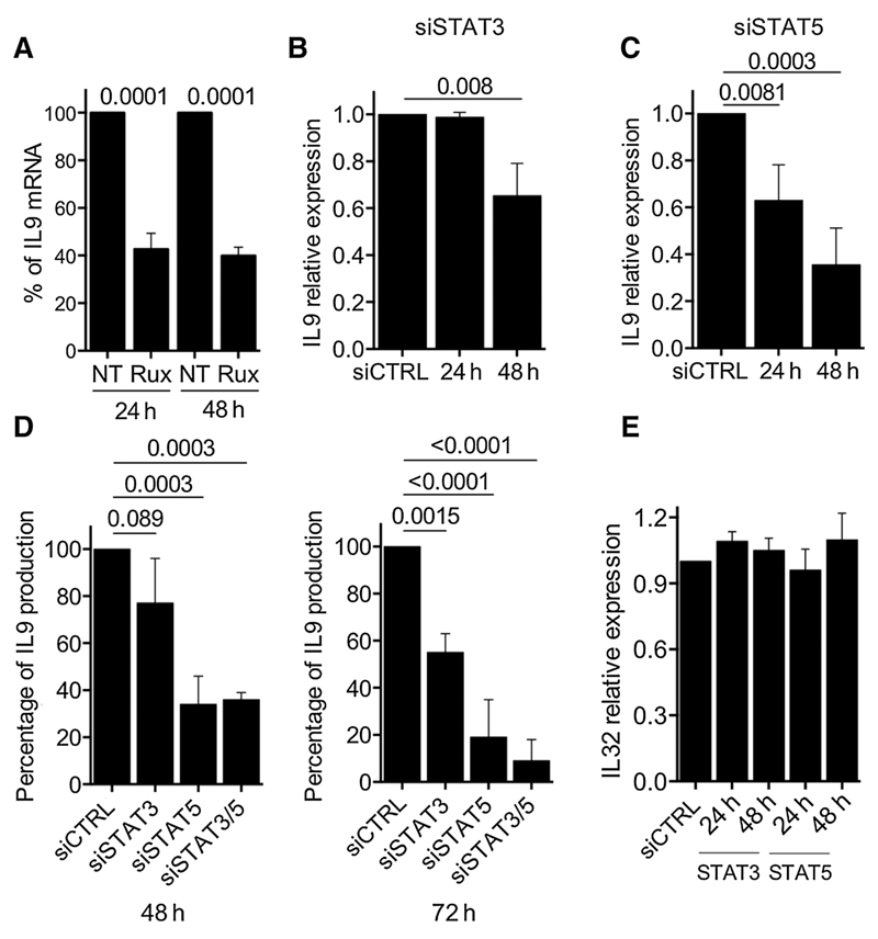
Results: Malignant and reactive T cells produce IL9 in lesional skin. Expression of the Th9 transcription factor IRF4 in malignant cells was heterogeneous, whereas reactive T cells expressed it uniformly. PUVA or UVB phototherapy diminished the frequencies of IL9- and IL9r-positive cells, as well as STAT3/5a and IRF4 expression in lesional skin. IL9 production was regulated by STAT3/5 and silencing of STAT5 or blockade of IL9 with neutralizing antibodies potentiated cell death after PUVA treatment in vitro IL9-depleted mice exhibited a reduction of tumor growth, higher frequencies of regulatory T cells, and activated CD4 and CD8 T lymphocytes.
Conclusions: Our results suggest that IL9 and its regulators are promising new targets for therapy development in mycosis fungoides. Clin Cancer Res; 22(13); 3328-39. ©2016 AACR.
©2016 American Association for Cancer Research.
Conflict of interest statement
No potential conflicts of interest were disclosed.
Figures
Similar articles
-
Jak3, STAT3, and STAT5 inhibit expression of miR-22, a novel tumor suppressor microRNA, in cutaneous T-Cell lymphoma.Oncotarget. 2015 Aug 21;6(24):20555-69. doi: 10.18632/oncotarget.4111. Oncotarget. 2015. PMID: 26244872 Free PMC article.
-
IL-15 and IL-17F are differentially regulated and expressed in mycosis fungoides (MF).Cell Cycle. 2014;13(8):1306-12. doi: 10.4161/cc.28256. Epub 2014 Mar 3. Cell Cycle. 2014. PMID: 24621498 Free PMC article.
-
Smad2/3 and IRF4 play a cooperative role in IL-9-producing T cell induction.J Immunol. 2013 Sep 1;191(5):2360-71. doi: 10.4049/jimmunol.1301276. Epub 2013 Aug 2. J Immunol. 2013. PMID: 23913959
-
Primary cutaneous T-cell lymphoma (mycosis fungoides and Sézary syndrome): part I. Diagnosis: clinical and histopathologic features and new molecular and biologic markers.J Am Acad Dermatol. 2014 Feb;70(2):205.e1-16; quiz 221-2. doi: 10.1016/j.jaad.2013.07.049. J Am Acad Dermatol. 2014. PMID: 24438969 Review.
-
Primary cutaneous T-cell lymphoma (mycosis fungoides and Sézary syndrome): part II. Prognosis, management, and future directions.J Am Acad Dermatol. 2014 Feb;70(2):223.e1-17; quiz 240-2. doi: 10.1016/j.jaad.2013.08.033. J Am Acad Dermatol. 2014. PMID: 24438970 Review.
Cited by
-
An overview of cutaneous T cell lymphomas.F1000Res. 2016 Jul 28;5:F1000 Faculty Rev-1882. doi: 10.12688/f1000research.8829.1. eCollection 2016. F1000Res. 2016. PMID: 27540476 Free PMC article. Review.
-
STAT3 Dysregulation in Mature T and NK Cell Lymphomas.Cancers (Basel). 2019 Nov 2;11(11):1711. doi: 10.3390/cancers11111711. Cancers (Basel). 2019. PMID: 31684088 Free PMC article. Review.
-
IL-9 and IL-9-producing cells in tumor immunity.Cell Commun Signal. 2020 Mar 30;18(1):50. doi: 10.1186/s12964-020-00538-5. Cell Commun Signal. 2020. PMID: 32228589 Free PMC article. Review.
-
Emerging drugs for the treatment of cutaneous T-cell lymphoma.Expert Opin Emerg Drugs. 2022 Mar;27(1):45-54. doi: 10.1080/14728214.2022.2049233. Epub 2022 Mar 8. Expert Opin Emerg Drugs. 2022. PMID: 35235473 Free PMC article. Review.
-
TH9 cells in skin disorders.Semin Immunopathol. 2017 Jan;39(1):47-54. doi: 10.1007/s00281-016-0607-8. Epub 2016 Nov 28. Semin Immunopathol. 2017. PMID: 27896634 Free PMC article. Review.
References
-
- Olsen E, Vonderheid E, Pimpinelli N, Willemze R, Kim Y, Knobler R, et al. Revisions to the staging and classification of mycosis fungoides and Sezary syndrome: a proposal of the International Society for Cutaneous Lymphomas (ISCL) and the cutaneous lymphoma task force of the European Organization of Research and Treatment of Cancer (EORTC) Blood. 2007;110:1713–22. - PubMed
-
- Ismail SA, Han R, Sanborn SL, Stevens SR, Cooper KD, Wood GS, et al. Immunohistochemical staining for CD45R isoforms in paraffin sections to diagnose mycosis fungoides-type cutaneous T-cell lymphoma. J Am Acad Dermatol. 2007;56:635–42. - PubMed
-
- Clark RA, Chong B, Mirchandani N, Brinster NK, Yamanaka K, Dowgiert RK, et al. The vast majority of CLA+ T cells are resident in normal skin. J Immunol. 2006;176:4431–9. - PubMed
Publication types
MeSH terms
Substances
Grants and funding
LinkOut - more resources
Full Text Sources
Other Literature Sources
Research Materials
Miscellaneous
