. 2016 Apr 1;412(1):99-113.
doi: 10.1016/j.ydbio.2016.01.036. Epub 2016 Feb 3.

UHRF1 regulation of Dnmt1 is required for pre-gastrula zebrafish development

Affiliations

UHRF1 regulation of Dnmt1 is required for pre-gastrula zebrafish development

Brandon Kent et al. Dev Biol. .

Abstract

Landmark epigenetic events underlie early embryonic development, yet how epigenetic modifiers are regulated to achieve rapid epigenome re-patterning is not known. Uhrf1 and DNA methyltransferase 1 (Dnmt1) are known to largely mediate maintenance DNA methylation and Uhrf1 is also required for both Dnmt1 localization and stability. Here, we investigate how these two key epigenetic modifiers regulate early zebrafish development and characterize the developmental consequences of disrupting their homeostatic relationship. Unlike Uhrf1 knockdown, which causes developmental arrest and death prior to gastrulation, overexpression of human UHRF1 (WT-UHRF1) caused asymmetric epiboly, inefficient gastrulation and multi-systemic defects. UHRF1 phosphorylation was previously demonstrated as essential for zebrafish embryogenesis, and we found that penetrance of the asymmetric epiboly phenotype was significantly increased in embryos injected with mRNA encoding non-phosphorylatable UHRF1 (UHRF1(S661A)). Surprisingly, both WT-UHRF1 and UHRF1(S661A) overexpression caused DNA hypomethylation. However, since other approaches that caused an equivalent degree of DNA hypomethylation did not cause the asymmetric epiboly phenotype, we conclude that bulk DNA methylation is not the primary mechanism. Instead, UHRF1(S661A) overexpression resulted in accumulation of Dnmt1 protein and the overexpression of both WT and a catalytically inactive Dnmt1 phenocopied the assymetric epiboly phenotype. Dnmt1 knockdown suppressed the phenotype caused by UHRF1(S661A) overexpression, and Uhrf1 knockdown suppressed the effect of Dnmt1 overexpression. Therefore, we conclude that the interaction between these two proteins is the mechanism underlying the gastrulation defects. This indicates that Dnmt1 stability requires UHRF1 phosphorylation and that crosstalk between the proteins is essential for the function of these two important epigenetic regulators during gastrulation.

Keywords: DNA methylation; Dnmt1; Epiboly; Epigenetics; Uhrf1; Zebrafish development.


Figures

Figure 1
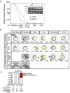
Figure 1. Distinct gastrulation defects in UHRF1 overexpressing embryos during early zebrafish embryogenesis
(A) Endogenous uhrf1 and dnmt1 are maternally provided. RT-qPCR and RT-PCR (inset, gel electrophoresis) display the relative transcript levels of uhrf1 in unfertilized eggs, shield stage embryos (6 hpf), prim 5 stage embryos (24 hpf), 3 dpf larvae, and 5 dpf larvae. (B) Progression of embryonic development in uninjected wild-type embryos and UHRF1 overexpressing embryos at each indicated stage. At high stage (3.3 hpf) all embryos appear normal and at 50% epiboly (~5.25 hpf) and shield stage (6.0 hpf), arrows indicate asymmetric epiboly and delay in dorsal organizer formation. At bud stage (10.0 hpf), arrows indicate failure to complete epiboly and premature development of emerging tail bud and at 7-8-Somites stage (~12.0 hpf) arrows depict a delay in somite formation, pinched off yolk and, in some cases, a lack of a developing tail. At Prim-5 stage (24.0 hpf) arrows indicate morphological defects such as under-developed eyes and head, and defects in tail extension. Scale bar = 500 micrometers. (C) The percent of embryos displaying asymmetric epiboly at 6 hpf in those injected with control RNA (EGFP and mpi) and WT-UHRF1. Significance of the percentage of the asymmetric epibly was determined by Fisher’s exact test. Asterisks (*) indicate p < 0.0001. The number of embryos and number of clutches for each condition is indicated.
Figure 2
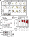
Figure 2. Overexpression of phosphorylation-deficient mutant UHRF1 (UHRF1S661A) phenocopies UHRF1 overexpressing embryos eliciting dramatic gastrulation defects
(A) Progression of embryonic development in uninjected and embryos injected with 100 pg of UHRF1S661A mRNA . At 50% epiboly (5.25 hpf), arrows indicate asymmetric epiboly. At Shield stage (6.0 hpf) arrows point to the missing shield and cells that did not undergo proper epiboly cell movements. At bud stage (10.0 hpf) arrows indicate failure to complete epiboly and premature development of tail bud. At 7-8-Somite stage (~12.0 hpf) arrows indicate disrupted anterior-posterior axis, somite formation, and irregular tail formation. At Prim-5 stage (24.0 hpf) arrows indicate defects in head and tail. Scale bar = 500 micrometers. (B) Curve displays the percent of embryos injected with 100 pg of WT-UHRF1 or UHRF1S661A or uninjected controls that were scored as normal; the stages set by the progression of uninjected control embryos (top) and hpf (bottom). Significance was determined by Log-rank Test (p < 0.0001). (C) Distribution of phenotypes of embryos injected with 25, 50 and 100 pg of WT-UHRF1 and UHRF1S661A mRNA. Penetrance of the asymmetric epiboly phenotype is statistically significant compared to uninjected controls at each indicated concentration by Fisher’s exact test. Asterisks (*) indicate p < 0.001. n.s. = not significant. Number of biological replicates (clutches), number of embryos per treatment, and percent of embryos displaying the asymmetric epiboly phenotype are indicated. (D) Western blot using the Myc tag to detect UHRF1 in WT-UHRF1 and UHRF1S661A overexpressing embryos. Mean levels of Myc-UHRF1 protein per treatment are quantified relative to tubulin and were determined to be equivalent by Student’s t-test (p = 0.36). n.s. = not significant.
Figure 3
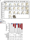
Figure 3. uhrf1 knockdown does not phenocopy UHRF1 overexpression
(A) Developmental progression of uninjected embryos and uhrf1 morphants (MO2-5’-UTR) at each indicated stage. At high stage (~3.3 hpf) both uninjected embryos and uhrf1 morphants appear normal and at 50% Epiboly (~5.25 hpf), arrow indicates arrest of uhrf1 morphant that have progressed to yolk extrusion. At shield stage (~6.0 hpf), arrow indicates significant developmental delay in uhrf1 morphants that did not arrest at high stage. Morphants that did arrest at high stage are dead by the time uninjected embryos have reached shield stage. At bud stage (~10.0 hpf), arrow indicates tail bud formation prior to the completion of epiboly in uhrf1 morphants. At 7-8 Somite stage (~12.0 hpf), arrows indicate disrupted CNS, somites and tail bud. At Prim 5 stage (~24.0 hpf), arrows indicate that all surviving uhrf1 morphants display CNS and tail defects. Scale bar = 500 micrometers. (B) Embryos were scored at 6 hpf for asymmetric epiboly, high stage arrest or delay. The depletion of endogenous Uhrf1 by injection of 13 ng 5’UTR uhrf1 morpholino does not affect the asymmetric epiboly phenotype caused by injection of 100 pg of WT-UHRF1 or UHRF1S661A mRNA. The co-injection of WT-UHRF1 or UHRF1S661A mRNA additively increases the percentage of the embryos affected by the asymmetric epiboly phenotype. The treatments, the number of biological replicates, number of embryos and the proportion of embryos displaying the asymmetric epiboly phenotype are indicated.
Figure 4
Figure 4. Knockdown of endogenous uhrf1 and overexpression of UHRF1 results in equivalent genome-wide DNA hypomethylation
Slot blot analysis and quantification of methylated genomic DNA of (A) embryos injected with 100 pg of WT-UHRF1 or UHRF1S661A mRNA, 3.4 ng of 5’UTR uhrf1 morpholino, and 0.43 pg of dnmt1 morpholino were normalized to their uninjected siblings at 3 hpf. (B) The same samples were assessed at 6 hpf, with the addition of samples exposed to 70 or 100 µM 5-AZA from 0.5 hpf normalized to uninjected, untreated siblings. Student’s t-test assessed determine significance between each respective treatment (p < 0.05). n.s. = not significant. Number of biological replicates are indicated.
Figure 5
Figure 5. dnmt1 knockdown does not phenocopy uhrf1 morphants or UHRF1 over-expressing embryos
(A) Developmental progression of uninjected embryos and those injected with 0.43 ng of dnmt1 morpholino at each indicated stage. Scale bar = 500 micrometers. (B) Quantification of developmental delay at 6 hpf in embryos injected with 0.43, 0.85, 1.28 and 1.7 ng of dnmt1 morpholino and uninjected control embryos. Fisher’s exact test was used to determine statistical significance. n.s. = not significant. (C) Representative western blot and quantification of Dnmt1 protein knockdown at indicated amount of dnmt1 morpholino. Student’s t-test was used to calculate the indicated p-values compared dnmt1 morpholino injected embryos to uninjected controls. Values are average of 3 biological replicates, error bars are standard deviation.
Figure 6
Figure 6. Dnmt1 protein levels are stabilized in UHRF1S661A over-expressing embryos
(A) dnmt1 mRNA expression at 6 hpf in uninjected embryos, WT-UHRF1 overexpressing embryos, and UHRF1S661A overexpressing embryos. Mean ΔCT values are indicated. Student’s t-test was used to determine p-value, n.s. indicates not significant (p< 0.05). (B) Western blot of Dnmt1, Myc-WT-UHRF1, and Myc-UHRF1S661A (top) and quantification of 5 biological replicates are shown with the median as the middle of the box and the span of the box indicating the 75th and 25th percentiles with the whiskers indicating the 90th and 10th percentiles. Student’s t-test was used to calculate p-values.
Figure 7
Figure 7. Dnmt1 overexpression phenocopies the asymmetric epiboly phenotype of UHRF1 and UHRF1S661A overexpressing embryos
(A) Developmental progression of uninjected and WT-dnmt1 overexpressing embryos at indicated stages. At High Stage (~3.3 hpf) all embryos appear normal at 50% epiboly (~5.25 hpf) and Shield stage (6.0 hpf) arrows indicate asymmetric epiboly and lack of dorsal organizer. At Bud stage (10.0 hpf) arrow points to the front of the migrating cells indicating delayed completion of epiboly. At Prim-5 stage (24.0 hpf) arrows indicate severe defects in head and tail with lack of head and tail extension. (B) Embryos were scored at 6 hpf for asymmetric epiboly and delay following injection of 1, 10, 12.5, 25, 50 or 100 pg of WT-dnmt1 mRNA (sense) or 25, 50, or 100 antisense dnmt1 RNA as a negative control. The number of clutches, total number of embryos scored and percent of asymmetric epiboly are indicated. (C) Overexpression of catalytically inactive dnmt1C1109S (MT-dnmt1) causes asymmetric epiboly. Asterisk indicates statistical significance (p < 0.05) using Fisher’s exact test. The number of embryos and biological replicates are indicated. n.s. = not significant.
Figure 8
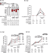
Figure 8. UHRF1 and Dnmt1 interact to cause asymmetric epiboly
(A) Embryos co-injected with 0.43 ng dnmt1 morpholino and either 100 pg WT-UHRF1 or UHRF1S661AmRNA were scored at 6 hpf for asymmetric epiboly. The number of biological replicates, number of embryos per treatment and the proportion of embryos with asymmetric epiboly are indicated. Significance was determined using Fisher’s exact test; asterisk indicates p < 0.001; n.s. = not significant. (B) Embryos from the same clutch were injected with 3.4 ng of 5’UTR uhrf1 MO or with 100 pg dnmt1 mRNA or a combination of these two and were scored at 6 hpf for asymmetric epiboly. The lines connect the data from each treatment in individual clutches. In all 4 replicates, co-injection suppressed the penetrance of the asymmetric epiboly phenotype. The number of biological replicates, the number of embryos per treatment, and the proportion of embryos displaying each phenotype are indicated. Significance of the asymmetric epiboly phenotype between the embryos injected with dnmt1 and the co-injected embryos was assessed by paired Students t-test. Embryos from the same clutch were injected with 25 pg of WT-dnmt1 mRNA with either 50 pg WT-UHRF1 (C) or UHRF1S661A (D) mRNA. Embryos were scored at 6 hpf for asymmetric epiboly. In all 6 replicates for WT-UHRF1 injection and 4 replicates for UHRF1S661A injection, co-injection enhanced the penetrance of the phenotype compared to each mRNA alone. Significance was determined using one-way ANOVA and p-values are indicated. The significance of the percentage of the increase of the asymmetric epiboly observed in the co-injected compared to the single injection treatment was assessed by paired t-test (p-values are indicated, n.s. indicates not significant).
Figure 9
Figure 9. Model of relationship between uhrf1 expression level dnmt1 expression level, DNA methylation, and progression through gastrulation
Knockdown of uhrf1 leads to genome-wide DNA hypomethylation and a pre-gastrula arrest phenotype (high stage arrest, 3.3 hpf). Overexpression of WT-UHRF1 and phospho-deficient UHRF1S661A also induces genome-wide DNA hypomethylation, but acting together with dnmt1, induces an asymmetric epiboly phenotype, ultimately disrupting gastrulation. This may be due in part to increased levels of non-phosphorylated UHRF1 and a subsequent increase in Dnmt1 protein resulting in the asymmetric epiboly phenotype due to a DNA methylation-independent function of Dnmt1.

Similar articles

Cited by

References

    1. Andersen IS, Reiner AH, Aanes H, Aleström P, Collas P. Developmental features of DNA methylation during activation of the embryonic zebrafish genome. Genome Biol. 2012;13:R65. - PMC - PubMed
    1. Anderson RM, Bosch JA, Goll MG, Hesselson D, Dong PDS, Shin D, Chi NC, Shin CH, Schlegel A, Halpern M, et al. Loss of Dnmt1 catalytic activity reveals multiple roles for DNA methylation during pancreas development and regeneration. Dev. Biol. 2009;334:213–223. - PMC - PubMed
    1. Arita K, Ariyoshi M, Tochio H, Nakamura Y, Shirakawa M. Recognition of hemi-methylated DNA by the SRA protein UHRF1 by a base-flipping mechanism. Nature. 2008;455:818–821. - PubMed
    1. Arita K, Isogai S, Oda T, Unoki M, Sugita K, Sekiyama N, Kuwata K, Hamamoto R, Tochio H, Sato M, et al. Recognition of modification status on a histone H3 tail by linked histone reader modules of the epigenetic regulator UHRF1. Proceedings of the National Academy of Sciences. 2012;109:12950–12955. - PMC - PubMed
    1. Avvakumov GV, Walker JR, Xue S, Li Y, Duan S, Bronner C, Arrowsmith CH, Dhe-Paganon S. Structural basis for recognition of hemi-methylated DNA by the SRA domain of human UHRF1. Nature. 2008;455:822–825. - PubMed

Publication types

MeSH terms

Substances