

Role of Intrinsic Protein Disorder in the Function and Interactions of the Transcriptional Coactivators CREB-binding Protein (CBP) and p300
- PMID: 26851278
- PMCID: PMC4807259
- DOI: 10.1074/jbc.R115.692020
Role of Intrinsic Protein Disorder in the Function and Interactions of the Transcriptional Coactivators CREB-binding Protein (CBP) and p300
Abstract
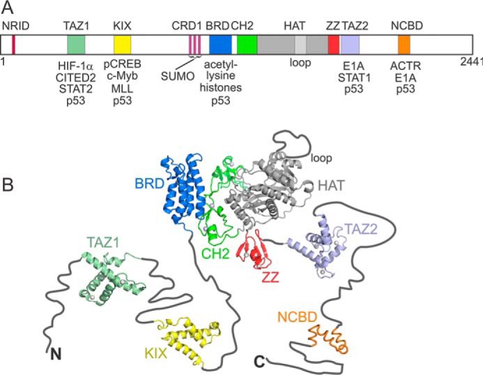
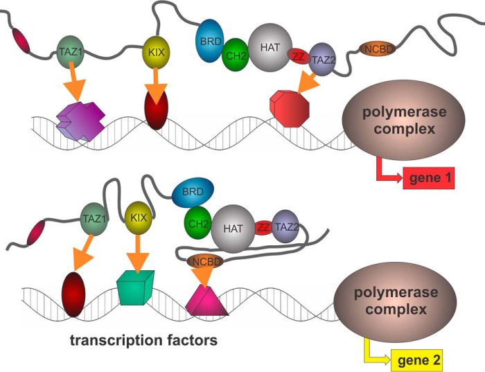
The transcriptional coactivators CREB-binding protein (CBP) and p300 undergo a particularly rich set of interactions with disordered and partly ordered partners, as a part of their ubiquitous role in facilitating transcription of genes. CBP and p300 contain a number of small structured domains that provide scaffolds for the interaction of disordered transactivation domains from a wide variety of partners, including p53, hypoxia-inducible factor 1α (HIF-1α), NF-κB, and STAT proteins, and are the targets for the interactions of disordered viral proteins that compete with cellular factors to disrupt signaling and subvert the cell cycle. The functional diversity of the CBP/p300 interactome provides an excellent example of the power of intrinsic disorder to facilitate the complexity of living systems.
Keywords: IDP; IDR; STAT transcription factor; cAMP response element-binding protein (CREB); coupled folding and binding; hypoxia-inducible factor (HIF); intrinsically disordered protein; intrinsically disordered region; protein-protein interaction; structure-function; transcriptional activation; transcriptional coactivator; viral oncoprotein.
© 2016 by The American Society for Biochemistry and Molecular Biology, Inc.
Figures
Similar articles
-
Complex regulation of the transactivation function of hypoxia-inducible factor-1 alpha by direct interaction with two distinct domains of the CREB-binding protein/p300.J Biol Chem. 2010 Jan 22;285(4):2601-9. doi: 10.1074/jbc.M109.021824. Epub 2009 Oct 30. J Biol Chem. 2010. PMID: 19880525 Free PMC article.
-
Mapping the interactions of adenoviral E1A proteins with the p160 nuclear receptor coactivator binding domain of CBP.Protein Sci. 2016 Dec;25(12):2256-2267. doi: 10.1002/pro.3059. Epub 2016 Oct 15. Protein Sci. 2016. PMID: 27699893 Free PMC article.
-
Conformational propensities of intrinsically disordered proteins influence the mechanism of binding and folding.Proc Natl Acad Sci U S A. 2015 Aug 4;112(31):9614-9. doi: 10.1073/pnas.1512799112. Epub 2015 Jul 20. Proc Natl Acad Sci U S A. 2015. PMID: 26195786 Free PMC article.
-
Transcriptional/epigenetic regulator CBP/p300 in tumorigenesis: structural and functional versatility in target recognition.Cell Mol Life Sci. 2013 Nov;70(21):3989-4008. doi: 10.1007/s00018-012-1254-4. Epub 2013 Jan 11. Cell Mol Life Sci. 2013. PMID: 23307074 Free PMC article. Review.
-
Modulating the masters: chemical tools to dissect CBP and p300 function.Curr Opin Chem Biol. 2018 Aug;45:195-203. doi: 10.1016/j.cbpa.2018.06.005. Epub 2018 Jul 17. Curr Opin Chem Biol. 2018. PMID: 30025258 Free PMC article. Review.
Cited by
-
Nuclear receptors in osteoclasts.Curr Opin Pharmacol. 2020 Aug;53:8-17. doi: 10.1016/j.coph.2020.03.002. Epub 2020 Jun 20. Curr Opin Pharmacol. 2020. PMID: 32569976 Free PMC article. Review.
-
Cyclic Adenosine Monophosphate: A Central Player in Gamete Development and Fertilization, and Possible Target for Infertility Therapies.Int J Mol Sci. 2022 Dec 1;23(23):15068. doi: 10.3390/ijms232315068. Int J Mol Sci. 2022. PMID: 36499392 Free PMC article.
-
Compendium of human transcription factor effector domains.Mol Cell. 2022 Feb 3;82(3):514-526. doi: 10.1016/j.molcel.2021.11.007. Epub 2021 Dec 3. Mol Cell. 2022. PMID: 34863368 Free PMC article. Review.
-
EP300 as a Molecular Integrator of Fibrotic Transcriptional Programs.Int J Mol Sci. 2023 Aug 1;24(15):12302. doi: 10.3390/ijms241512302. Int J Mol Sci. 2023. PMID: 37569677 Free PMC article. Review.
-
Role of Backbone Dynamics in Modulating the Interactions of Disordered Ligands with the TAZ1 Domain of the CREB-Binding Protein.Biochemistry. 2019 Mar 12;58(10):1354-1362. doi: 10.1021/acs.biochem.8b01290. Epub 2019 Feb 22. Biochemistry. 2019. PMID: 30775911 Free PMC article.
References
-
- Minezaki Y., Homma K., Kinjo A. R., and Nishikawa K. (2006) Human transcription factors contain a high fraction of intrinsically disordered regions essential for transcriptional regulation. J. Mol. Biol. 359, 1137–1149 - PubMed
-
- Sigler P. B. (1988) Transcriptional activation: acid blobs and negative noodles. Nature 333, 210–212 - PubMed
-
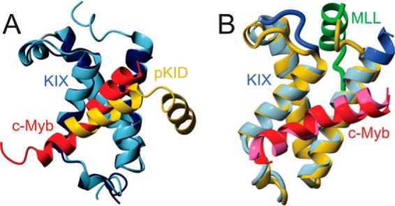
- Radhakrishnan I., Pérez-Alvarado G. C., Parker D., Dyson H. J., Montminy M. R., and Wright P. E. (1997) Solution structure of the KIX domain of CBP bound to the transactivation domain of CREB: a model for activator:coactivator interactions. Cell 91, 741–752 - PubMed
-
- Wright P. E., and Dyson H. J. (1999) Intrinsically unstructured proteins: re-assessing the protein structure-function paradigm. J. Mol. Biol. 293, 321–331 - PubMed
Publication types
MeSH terms
Substances
Associated data
- Actions
- Actions
Grants and funding
LinkOut - more resources
Full Text Sources
Other Literature Sources
Research Materials
Miscellaneous
