Review
. 2016 Mar 25;291(13):6696-705.
doi: 10.1074/jbc.R115.695056. Epub 2016 Feb 5.

Modulation of Intrinsically Disordered Protein Function by Post-translational Modifications

Affiliations
Review

Modulation of Intrinsically Disordered Protein Function by Post-translational Modifications

Alaji Bah et al. J Biol Chem. .

Abstract

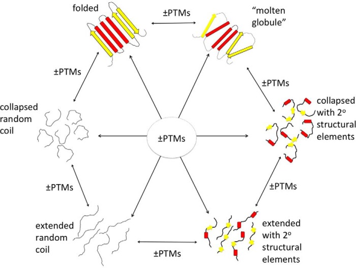
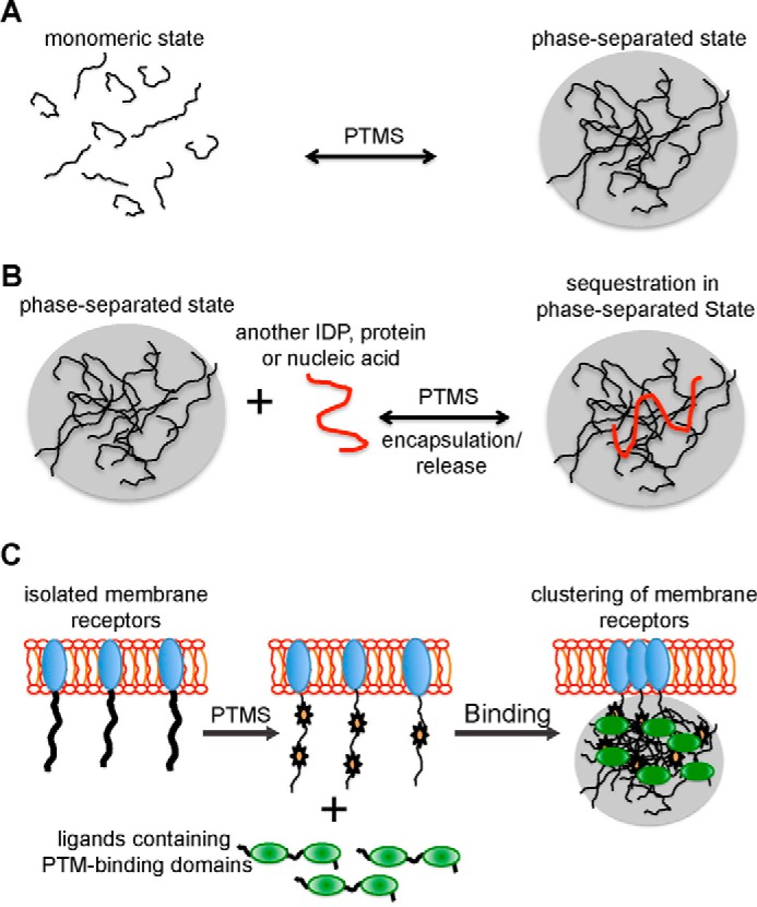
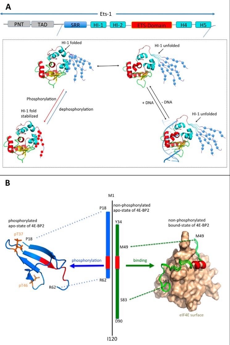
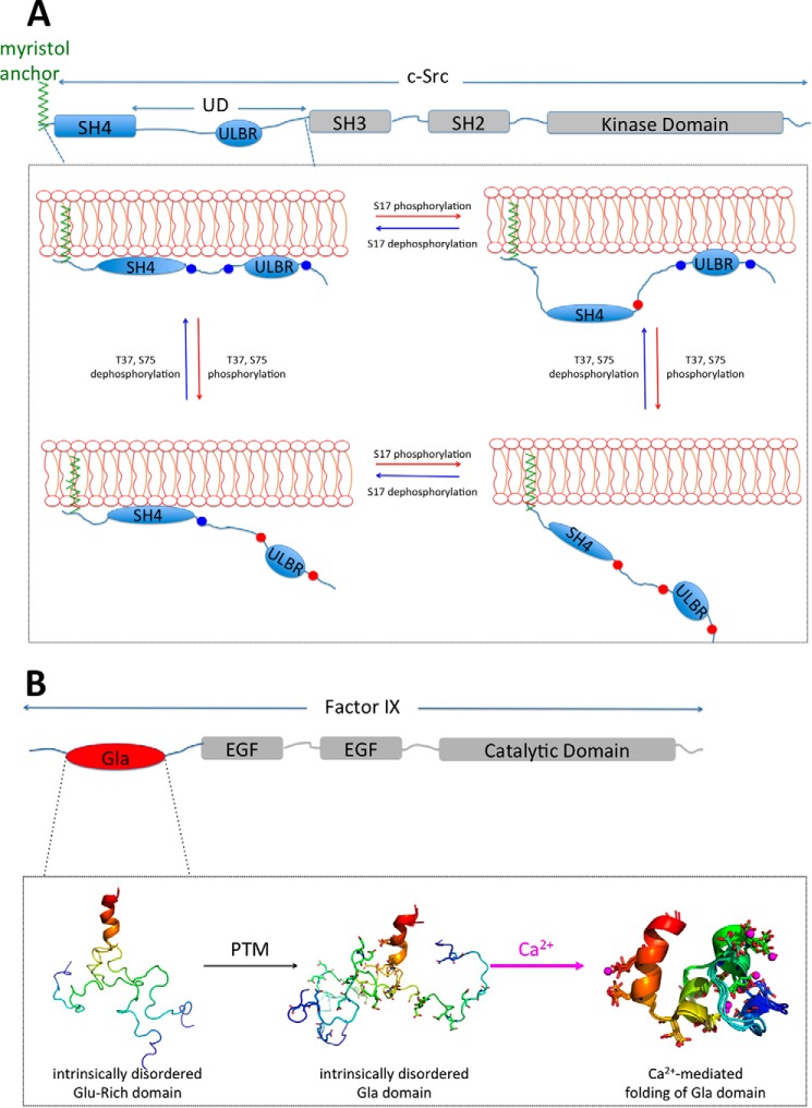
Post-translational modifications (PTMs) produce significant changes in the structural properties of intrinsically disordered proteins (IDPs) by affecting their energy landscapes. PTMs can induce a range of effects, from local stabilization or destabilization of transient secondary structure to global disorder-to-order transitions, potentially driving complete state changes between intrinsically disordered and folded states or dispersed monomeric and phase-separated states. Here, we discuss diverse biological processes that are dependent on PTM regulation of IDPs. We also present recent tools for generating homogenously modified IDPs for studies of PTM-mediated IDP regulatory mechanisms.

Keywords: intrinsically disordered protein; post-translational modification (PTM); protein conformation; protein-DNA interaction; protein-protein interaction; regulation.


Figures

FIGURE 1.
FIGURE 1.
Post-translational modifications of IDPs can induce diverse structural changes. PTM-mediated structural changes within IDP ensembles and between IDPs and folded states are shown. These conformational ensembles range from folded and molten globules to extended and collapsed disordered ensembles (represented with six members) with and without transient or stable secondary structural elements.
FIGURE 2.
FIGURE 2.
IDPs can form PTM-induced phase-separated droplets in cellular organization such as membraneless organelles and signaling puncta. A and B, PTMs can enhance the assembly or disassembly of the phase-separated state (A) or control whether another molecule can be encapsulated into or released from the droplet (B). C, weak multivalent interactions between IDPs with multiple PTM sites and proteins containing multiple PTM binding modules can also result in phase separation to form cellular bodies or signaling puncta.
FIGURE 3.
FIGURE 3.
IDP-mediated disorder-to-order transitions to regulate protein complex formation. A, top, schematic representation of Ets-1, composed of an N-terminal protein interaction domain (PNT, gray), the transactivation domain (TAD, gray) followed by a C-terminal SRR (blue), and the IM domain (cyan) flanking the ETS DNA binding domain (red/yellow). Lower, in the apo state, helix HI-1 of the IM domain is in dynamic equilibrium between folded and disordered conformations (Protein Data Bank (PDB) codes 1R36 (65) and 1MDM (64), respectively). Upon multi-site phosphorylation on the SRR (blue to red circles), SRR dynamic fluctuations are damped, enhancing transient stabilizing interactions with the ETS and IM domains, further reducing DNA binding. B, regulation of the 4E-BP2·eIF4E complex by phosphorylation of Thr37 (pT37) and Thr46 (pT46) (orange stick model) leads to folding of residues Pro18–Arg62 (blue, PDB code: 2MX4), with further phosphorylation at Ser65, Thr70, and Ser83 stabilizing the folded state, enabling translation initiation. In the absence of eIF4E, non-phosphorylated 4E-BP2 (black) is disordered albeit with significant transient secondary structural elements (69). 4E-BP2 utilizes residues from about Tyr34 to Asp90 (green) consisting of a helical element (red) containing the canonical binding (54YXXXX60) motif and a flexible secondary binding site involving 78IPGVT82 for eIF4E (brown surface) binding. (Note: The longest observed fragment of a 4E-BP in complex with eIF4E in a crystal structure (72) is only Met49 to Ser82 (PDB code: 4UED).) This dynamic complex is represented by an ensemble of three conformers of 4E-BP2 on the surface of eIF4E.
FIGURE 4.
FIGURE 4.
PTM-regulated docking of IDRs onto a phospholipid membrane. A, anchoring of c-Src via SH4 and UD (77). Top, schematic representation of the modular domains of c-Src showing the IDRs SH4 and UD (blue) as well as the SH3 and SH2 interaction domains and a C-terminal tyrosine kinase domain (gray). In addition to the myristoyl group (green) attached to Gly2 of the SH4 domain, the SH4 domain and UD can also bind phospholipids. Phosphorylation of the SH4 at Ser17 by PKA and of the UD at Thr37/Ser75 by cyclin-dependent kinase (CDK) controls the SH4 and Unique lipid binding region (ULBR) localization to the phospholipid membrane. B, γ-carboxylation of the Gla domain enables the calcium-mediated disorder-to-folded transition of the Gla domain. Top, schematic representation of factor IX with the Gla domain (red) followed by two tandem EGF domains and a C-terminal serine protease domain (gray). The N-terminal Glu-rich IDR undergoes multisite γ-carboxylation to form the Gla domain (Gla residues shown in stick representation, PDB code: 1CFH). Upon Ca2+ binding, the entire Gla domain folds into a four-helix bundle (calcium ions in magenta spheres, PDB codes: 1J35, 1CFI), which can bind phospholipid membranes. The Gla domain is represented by an ensemble of three conformers each color-coded N- to C-terminal from blue to red, respectively, with the C-terminal helix superimposed.

Similar articles

Cited by

References

    1. Dyson H. J., and Wright P. E. (2005) Intrinsically unstructured proteins and their functions. Nat. Rev. Mol. Cell Biol. 6, 197–208 - PubMed
    1. van der Lee R., Buljan M., Lang B., Weatheritt R. J., Daughdrill G. W., Dunker A. K., Fuxreiter M., Gough J., Gsponer J., Jones D. T., Kim P. M., Kriwacki R. W., Oldfield C. J., Pappu R. V., Tompa P., Uversky V. N., Wright P. E., and Babu M. M. (2014) Classification of intrinsically disordered regions and proteins. Chem. Rev. 114, 6589–6631 - PMC - PubMed
    1. Xie H., Vucetic S., Iakoucheva L. M., Oldfield C. J., Dunker A. K., Obradovic Z., and Uversky V. N. (2007) Functional anthology of intrinsic disorder. 3. Ligands, post-translational modifications, and diseases associated with intrinsically disordered proteins. J. Proteome Res. 6, 1917–1932 - PMC - PubMed
    1. Wright P. E., and Dyson H. J. (2009) Linking folding and binding. Curr. Opin. Struct. Biol. 19, 31–38 - PMC - PubMed
    1. Dosztányi Z., Chen J., Dunker A. K., Simon I., and Tompa P. (2006) Disorder and sequence repeats in hub proteins and their implications for network evolution. J. Proteome Res. 5, 2985–2995 - PubMed

Publication types

MeSH terms

LinkOut - more resources