Review
. 2016 Mar 25;291(13):6706-13.
doi: 10.1074/jbc.R115.692590. Epub 2016 Feb 5.

Expanding the Range of Protein Function at the Far End of the Order-Structure Continuum

Affiliations
Review

Expanding the Range of Protein Function at the Far End of the Order-Structure Continuum

Virginia M Burger et al. J Biol Chem. .

Abstract

The traditional view of the structure-function paradigm is that a protein's function is inextricably linked to a well defined, three-dimensional structure, which is determined by the protein's primary amino acid sequence. However, it is now accepted that a number of proteins do not adopt a unique tertiary structure in solution and that some degree of disorder is required for many proteins to perform their prescribed functions. In this review, we highlight how a number of protein functions are facilitated by intrinsic disorder and introduce a new protein structure taxonomy that is based on quantifiable metrics of a protein's disorder.

Keywords: biophysics; conformational change; intrinsically disordered protein; protein folding; protein structure.


Figures

FIGURE 1.
FIGURE 1.
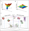
Order and structure in proteins. A and B, schematic of an energy landscape for an ordered protein (A) and a disordered protein (B). Red tones indicate relatively high energy conformations, and blue tones indicate low energy conformations. C, order-structure continuum. Proteins with varying degrees of order and secondary structure propensity are shown in their approximate position on an order-structure continuum. Helices are shown in red, β-strands are shown in purple, and turns and unstructured regions are shown in green. Red, purple, and blue lines are used to indicate location along helical content, β-strand content, and order axes in three dimensions. Protein Data Bank (PDB) IDs left to right are: 2k3j (71), 1bh4 (72), PED Entry 9AAA (73), 1k7b (74), PED Entry 7AAA (75), 2kac (76), 2k3g (77), and 1qjp (78). PED refers to the Protein Ensemble Database (79).
FIGURE 2.
FIGURE 2.
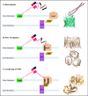
Roles of colicin E9's intrinsically unstructured region within its T-domain (IUTD). In each panel, the domains of ColE9 are shown in pink tones: R, pale pink; C, red; T, carnation pink. The immunity protein, Im9, which inhibits the cytotoxic function of ColE9 in the extracellular space, is shown in blue bound to C. The ColE9-binding sites for OmpF are shown as black rectangles, and the binding site for TolB is shown as a green rectangle. A, after binding BtuB, the BtuB-ColE9 complex diffuses along the membrane to locate and bind OmpF, aided by the extended search radius provided by the IUTD. A structure of BtuB bound to the receptor domain from the similar protein ColE3 is shown to the right (PDB ID: 1ujw (80)). B, the IUTD forms an initial complex with one OmpF pore. Side and top views of IUTD residues 2–16 bound to OmpF are shown to the right (PDB ID: 3O0e chains A, C, E, and L (33)). C, the IUTD passes further through OmpF into the periplasm and weaves back into OmpF, binding OmpF in two pores. The TolB-binding site is now exposed to the periplasm, allowing it to bind TolB, which in turn binds TolA, forming the translocon. The structure of the TBE bound to TolB is shown to the right (PDB ID: 2ivz chains D and H (37)).
FIGURE 3.
FIGURE 3.
The 4E-BP2 protein (green, with the primary eIF4E-binding site shown in violet) is disordered in its unphosphorylated, unbound state. Upon binding eIF4E (shown in white), its primary binding site adopts a helical conformation, but the remaining residues remain largely disordered and exposed for phosphorylation (PDB ID: 3am7, 4E-BP2 residues 47–65). Phosphorylation of 4E-BP2 causes it to fold into a binding-incompetent β-strand structure (PDB ID: 2mx4, 4E-BP2 residues 47–62 (41)). The disordered ensemble was generated with Mollack with chemical shift data from Biological Magnetic Resonance Bank (BMRB) 19114 for non-phosphorylated 4E-BP2 residues 1–120 with an N-terminal MPLGSPEF tag (46).
FIGURE 4.
FIGURE 4.
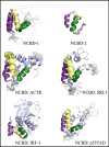
Conformations of NCBD. Available NMR and x-ray structures of NCBD are shown in complex with different binding partners or in isolation, aligned according to helix 1. NCBD is colored white, with helix 1 in purple, helix 2 in yellow, and helix 3 in green. Binding partners are shown in blue. For visibility of NMR models, only one conformation of each binding partner is shown, whereas all NCBD conformations are shown. Clockwise from upper left, the PDB IDs are 2kkj (54), 1jjs (50), 2c52 (82), 2l14 (84), 1zoq (83), and 1kbh (53). Figures were made in PyMOL (81).

Similar articles

Cited by

References

    1. McCammon J. A., Gelin B. R., and Karplus M. (1977) Dynamics of folded proteins. Nature 267, 585–590 - PubMed
    1. Wright P. E., and Dyson H. J. (1999) Intrinsically unstructured proteins: re-assessing the protein structure-function paradigm. J. Mol. Biol. 293, 321–331 - PubMed
    1. Fisher C. K., and Stultz C. M. (2011) Protein structure along the order-disorder continuum. J. Am. Chem. Soc. 133, 10022–10025 - PMC - PubMed
    1. Uversky V. N. (2013) A decade and a half of protein intrinsic disorder: biology still waits for physics. Protein Sci. 22, 693–724 - PMC - PubMed
    1. Marsh J. A., Teichmann S. A., and Forman-Kay J. D. (2012) Probing the diverse landscape of protein flexibility and binding. Curr. Opin Struct. Biol. 22, 643–650 - PubMed

Publication types

MeSH terms

LinkOut - more resources