. 2016 Apr:431:22-30.
doi: 10.1016/j.jim.2016.02.003. Epub 2016 Feb 3.

pMINERVA: A donor-acceptor system for the in vivo recombineering of scFv into IgG molecules

Affiliations

pMINERVA: A donor-acceptor system for the in vivo recombineering of scFv into IgG molecules

M Batonick et al. J Immunol Methods. 2016 Apr.

Abstract

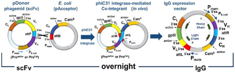
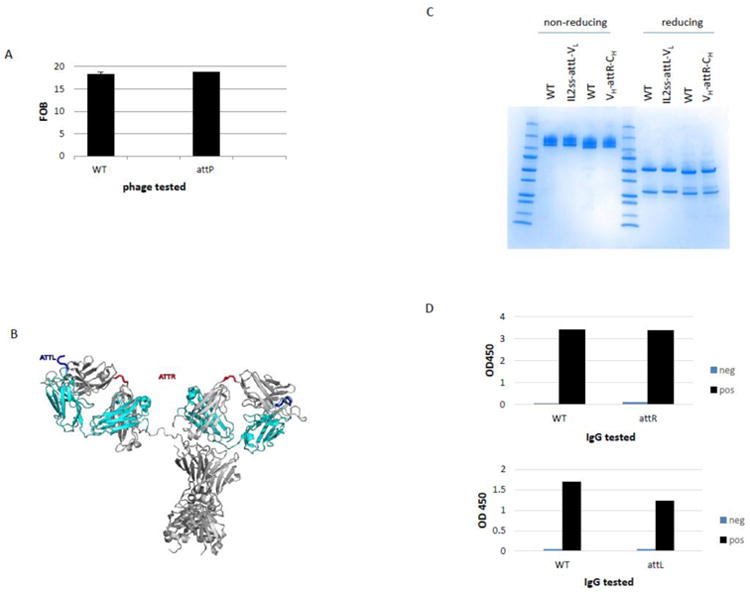
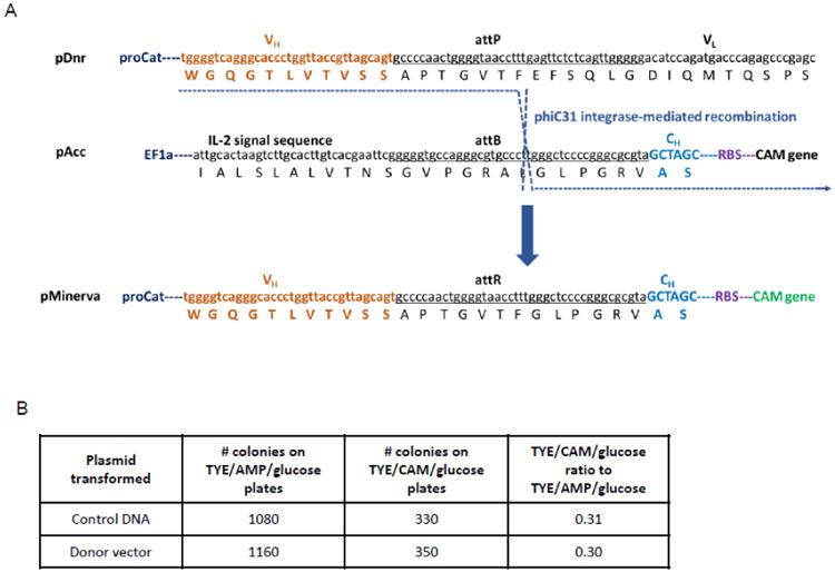
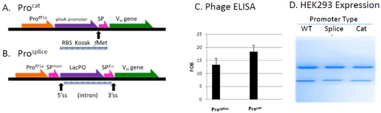
Phage display is the most widely used method for selecting binding molecules from recombinant antibody libraries. However, validation of the phage antibodies often requires early production of the cognate full-length immunoglobulin G (IgG). The conversion of phage library outputs to a full immunoglobulin via standard subcloning is time-consuming and limits the number of clones that can be evaluated. We have developed a novel system to convert scFvs from a phage display vector directly into IgGs without any in vitro subcloning steps. This new vector system, named pMINERVA, makes clever use of site-specific bacteriophage integrases that are expressed in Escherichia coli and intron splicing that occurs within mammalian cells. Using this system, a phage display vector contains both bacterial and mammalian regulatory regions that support antibody expression in E. coli and mammalian cells. A single-chain variable fragment (scFv) antibody is expressed on the surface of bacteriophage M13 as a genetic fusion to the gpIII coat protein. The scFv is converted to an IgG that can be expressed in mammalian cells by transducing a second E. coli strain. In that strain, the phiC31 recombinase fuses the heavy chain constant domain from an acceptor plasmid to the heavy chain variable domain and introduces controlling elements upstream of the light chain variable domain. Splicing in mammalian cells removes a synthetic intron containing the M13 gpIII gene to produce the fusion of the light chain variable domain to the constant domain. We show that phage displaying a scFv and recombinant IgGs generated using this system are expressed at wild-type levels and retain normal function. Use of the pMINERVA completely eliminates the labor-intensive subcloning and DNA sequence confirmation steps currently needed to convert a scFv into a functional IgG Ab.

Keywords: IgG; Recombineering; ScFv; Splicing.


Figures

Fig. 1
Fig. 1. Description of the pMINERVA transformer system
The scFv Abs encoded on the pDonor vector as M13gpIII-fusions can be screened in a phage display biopanning procedure to identify a phagemid encoding a scFv with the desired biophysical properties. This phagemid is transduced into an E. coli strain expressing phiC31 integrase and harboring an IgG acceptor vector. The product of the recombination event introduces a polyadenylation signal site adjacent to the 3′ end of the CH gene. Furthermore, the recombination event introduces both a mammalian promoter and functional protein initiation site 5′ to the VL gene. Of special note, the linker between the VH and VL domains of the scFv is composed of a phiC31 36-bp attP site that is able to function as both a: i) peptide linker between the heavy and light variable domains, and ii) 36-bp functional substrate for phiC31 integrase. [Pmam, mammalian promoter; 5′ss and 3′ss, splice signals; VL, variable section of the light chain; VH, variable section of the heavy chain; gp3, phage M13 gene3 product; PE.coli, E.coli promoter; CH or Fc, constant region of the heavy chain; attB, attP, substrates for an integrase gene; attR and attL, products of an integrase gene; polyA, polyadenylation sequence; CamS, CamR, chloramphenicol resistance gene without and with a promoter, respectively; Prosplice, Procat, dual-function promoter-types (see text and Fig 5 for details); RBS, ribosome binding site].
Fig. 2
Fig. 2. Expression and function of scFv and IgGs containing phiC31 integrase sites
(A) The same scFv with two different linker sequences, WT (Gly4Ser)3 or the phiC31 attP site in reading frame 2, was produced. Both phage-scFvs were tested in an ELISA against the purified target protein or a non-relevant antigen control. Anti-M13 antibody that is conjugated to Horseradish peroxidase (HRP) was used for phage detection and the enzyme linked immunosorbent assay (ELISA) was developed with Ultra Tetramethylbenzidine (TMB) reagent. The fold over background (FOB), which is the signal against target over the signal against non-relevant control, is shown for each phage tested. Error bars represent the standard deviation of phage binding tested in triplicate (B) IgG molecule modeled with attL and attR. The attL (thick blue loop) and attR (thick red loop) peptides are inserted schematically in a typical human IgG1 molecule (PDB ID: 1hzh) shown as ribbons (heavy chain: white, light chain: cyan). (C) The same IgG, with either no linker or the recombined phiC31 integrase attL site between the IL2 signal sequence (ss) and VL, was produced. In parallel, expression of the wild-type IgG was compared to expression of the same IgG with the recombined phiC31 integrase site attR site between VH and CH. A coommassie stained SDS-PAGE gel is shown. (D) An IgG with attL between the IL2ss and the VL (top graph) and an IgG with attR between VH and CH (bottom graph) were tested for binding to both a specific and a non-relevant target antigen in a cell ELISA. The binding of both molecules was compared to the binding of a wildtype IgG. Both ELISAs used anti-human-HRP to detect the IgGs and the ELISA was developed with Ultra TMB reagent. The ODs at 450nm are shown.
Fig. 3
Fig. 3. Positive selection of phiC31 integrase activity
(A) Two plasmids pDonor and pAcceptor, each having a needed component in trans for a functional camR gene were constructed. The attP site (underlined) in pDonor was flanked upstream by the E. coli 5′ controlling elements and. The attB site (lower case DNA sequence) in pAcceptor was placed 5‟ of the promoter-less camR gene. Successful recombination between the attP and attB sites on the two plasmids in the presence of the phiC31 integrase (blue dashed line) generates the co-integrant (pMINERVA; bottom sequence) and fuses an E. coli promoter in front of the bicistronic heavy chain-CamR message. (B) Competent TG1 cells containing pAcceptor were transformed with pDonor or a control mock-recombined vector and grown on plates containing ampicillin or chloramphenicol. The ratio of colonies on the ampicillin plates to the chloramphenicol plates was calculated.
Fig. 4
Fig. 4. Expression and function of scFv and IgGs containing splice sites flanking the M13 gpIII gene
(A) Phage-scFv fusions with and without splice sites flanking the M13 gpIII gene in pDonor were produced from E.coli and tested for functionality in a phage ELISA. The phage-scFv were tested for binding to purified target antigen and a non-relevant control protein. The fold over background (FOB) is shown for both and error bars represent the standard deviation of phage binding tested in triplicate (B) The same IgG, with either no linker or the intron containing the M13 gpIII gene between the VH and CH was produced. An ochre stop codon placed 3′ of the M13 gpIII gene prevents full length light chain protein expression from non-spliced mRNAs. Upper arrow indicates the band corresponding to the heavy chain and the lower arrow corresponds to the light chain gene product in the SDS-PAGE. IgG molecules were tested for functionality in an ELISA using purified antigen. FOB (fold-over-background) signal is shown for both.
Fig. 5
Fig. 5. Dual expression promoter systems
(A) Promoter type Procat. In the catenated promoter system, the ATGs downstream of the mammalian EF1a promoter (PEF1a) are removed from the downstream polyhedron (not shown) and PhoA bacterial promoter. The Kozak sequence and E.coli ribosomal binding site (RBS) are designed such that the first ATG fMet start site for either bacterial, insect (not shown) or mammalian expression is identical. In this case, the same signal sequence (SigPep) is used for all three organisms. (B) Promoter type Prosplice. In this dual function promoter, the LacPO and bacterial signal peptide (SigPepE.c.) sequence are contained within the mammalian intron. The bacterial signal peptide sequence overlaps the 3′ splice site (3′ss). In E. coli, transcription from the bacterial promoter within the mammalian intron results in the expression of the scFv in the bacterial periplasm fused to the M13 gpIII protein in an amber-suppressing strain of E.coli (for example, TG1). In mammalian cells, intron splicing of the mRNA at the 5′ (5′ss) and 3′ss removes the bacterial LacPO regulatory sequences located within the intron. Intron splicing generates the mammalian signal sequence. (C) Phage-scFv production from Procat and Prosplice promoters. Phage were tested for binding to purified target antigen and to a non-relevant control antigen in a phage ELISA. FOB (fold-over-background) signal is shown and error bars represent the standard deviation of phage binding tested in triplicate (D) Expression of the wild type (wt), Prosplice and Procat promoters in HEK293 mammalian cells. IgG purification from HEK293 cells where the IgG heavy chain gene was under the expression control of either an EF1A promoter alone, Prosplice or Procat.

Similar articles

Cited by

References

    1. Prinz F, Schlange T, Asadullah K. Believe it or not: how much can we rely on published data on potential drug targets? Nat Rev Drug Discov. 2011;10:712. - PubMed
    1. Bradbury A, Pluckthun A. Reproducibility: Standardize antibodies used in research. Nature. 2015;518:27–29. - PubMed
    1. Bradbury AM, Pluckthun A. Antibodies: validate recombinants once. Nature. 2015;520:295. - PubMed
    1. Miersch S, Li Z, Hanna R, McLaughlin ME, Hornsby M, Matsuguchi T, Paduch M, Saaf A, Wells J, Koide S, Kossiakoff A, Sidhu SS. Scalable high throughput selection from phage-displayed synthetic antibody libraries. J Vis Exp. 2015 doi: 10.3791/51492:51492. - DOI - PMC - PubMed
    1. Taussig MJ, Schmidt R, Cook EA, Stoevesandt O. Development of proteome-wide binding reagents for research and diagnostics. Proteomics Clin Appl. 2013;7:756–766. - PubMed

Publication types

MeSH terms