Review
. 2016 Mar:51:92-105.
doi: 10.1016/j.semcdb.2016.02.001. Epub 2016 Feb 3.

Frogs as integrative models for understanding digestive organ development and evolution

Affiliations
Review

Frogs as integrative models for understanding digestive organ development and evolution

Mandy Womble et al. Semin Cell Dev Biol. 2016 Mar.

Abstract

The digestive system comprises numerous cells, tissues and organs that are essential for the proper assimilation of nutrients and energy. Many aspects of digestive organ function are highly conserved among vertebrates, yet the final anatomical configuration of the gut varies widely between species, especially those with different diets. Improved understanding of the complex molecular and cellular events that orchestrate digestive organ development is pertinent to many areas of biology and medicine, including the regeneration or replacement of diseased organs, the etiology of digestive organ birth defects, and the evolution of specialized features of digestive anatomy. In this review, we highlight specific examples of how investigations using Xenopus laevis frog embryos have revealed insight into the molecular and cellular dynamics of digestive organ patterning and morphogenesis that would have been difficult to obtain in other animal models. Additionally, we discuss recent studies of gut development in non-model frog species with unique feeding strategies, such as Lepidobatrachus laevis and Eleutherodactylous coqui, which are beginning to provide glimpses of the evolutionary mechanisms that may generate morphological variation in the digestive tract. The unparalleled experimental versatility of frog embryos make them excellent, integrative models for studying digestive organ development across multiple disciplines.

Keywords: Eleutherodactylous coqui; Embryo; Evolution; Gut; Lepidobatrachus; Morphogenesis; Specification; Xenopus.


Figures

Figure 1
Figure 1. Wnt, RA, and FGF pattern the foregut
The primitive gut tube is regionalized along both A–P and D–V axes. During gastrulation, RA (red) is required for dorsal pancreas (DP; purple) specification, likely by inhibiting Shh expression. Slightly later, the foregut is distinguished from the hindgut by a gradient of Wnt signaling (orange). High posterior Wnt specifies the hindgut domain, while low anterior Wnt (yellow; limited by Sfrp5) signals through the Fzd7 receptor to promote foregut fates and initiate cellular morphogenesis. Finally, a gradient of FGF signaling from the neighboring cardiac/lateral plate mesoderm segregates ventral foregut organs; prolonged, higher levels of FGF are needed to specify liver (green) versus ventral pancreas (VP; blue).
Figure 2
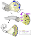
Figure 2. Intestine lengthening involves Hedghog- and Wnt/PCP-mediated endoderm cell polarization, rearrangement and epithelial differentiation
Initially, the endoderm cells of the prospective intestine are rounded, unpolarized and disorganized. Signaling via Hedgehogs (HH; from the endoderm) induces foxF1 expression in the surrounding mesoderm layer of the gut tube. This facilitates reciprocal signaling from the now differentiating visceral mesoderm, which regulates the rate of epithelial differentiation in the underlying endoderm. Concomitant non-canonical Wnt signals (presumably from the mesoderm) are required for the endoderm cells to become polarized, starting with the outermost (most basal) layers and progressing towards the center of the gut tube. Both actomyosin contractile forces, regulated by ROCK, and microtubule organization, regulated by JNK, are required to dynamically remodel adhesive contacts between the polarized endoderm cells. This enables productive radial intercalation of the most central cells into the outermost layer, resulting in tissue lengthening and the morphogenesis of a single layer intestinal epithelium.
Figure 3
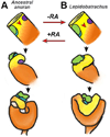
Figure 3. Altered RA signaling may have led to a novel foregut morphology
A) In the hypothetical ancestral anuran (represented by Xenopus), the herbivorous tadpole requires only a rudimentary stomach. The foregut domain of the primitive gut tube is small relative to the hindgut domain, causing the gastroduodenal (GD) loop to form in a relatively anterior position and acquire an acute curvature during later foregut morphogenesis. B) In contrast, in the carnivorous Lepidobatrachus tadpole, which requires a capacious stomach, the ratio of foregut to hindgut is greater, and the GD loop forms in a more posterior position. Consequently, the larger carnivore stomach becomes more transversely oriented. This anatomical change may have been dependent on a decrease in RA signaling during foregut development in the Lepidobatrachus lineage, since inhibiting RA in Xenopus (representative of the ancestral condition) transforms the GD loop to resemble that observed in Lepidobatrachus. Conversely, exposing Lepidobatrachus embryos to excess RA elicits a more typical foregut configuration.
Figure 4
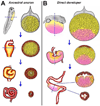
Figure 4. Endoderm morphogenesis in ancestral versus direct-developing frog species
A) In ancestral frogs that produce feeding (exotrophic) tadpoles, all of the yolky vegetal endoderm cells (yellow) in the primitive gut tube (PGT) are used to generate the lining of the tadpole gut. As these cells radially rearrange (see Figure 2) and differentiate into the final digestive epithelium (orange/red), a central lumen is formed and the intestine is lengthened to form a long, coiled tract. The extensive gut is eventually remodeled to a shorter adult tract during metamorphosis (not shown). B) In contrast, in the direct-developing (endotrophic) frog embryo, a subset of the vegetal endoderm cells are fated to become nutritional endoderm (NE; pink), a cell type that does not rearrange nor contribute to the gut epithelium. Instead, these cells are gradually depleted of their yolk, extruded and eliminated as waste. Consequently, the PGT does not generate a long tract, and the developing froglet directly forms a short adult-length intestine.

Similar articles

Cited by

References

    1. Keller R. Cell migration during gastrulation. Current opinion in cell biology. 2005;17:533–541. - PubMed
    1. Keller R, Davidson LA, Shook DR. How we are shaped: the biomechanics of gastrulation. Differentiation. 2003;71:171–205. - PubMed
    1. Spemann H, Mangold H. Induction of embryonic primordia by implantation of organizers from a different species. 1923. Int J Dev Biol. 2001;45:13–38. - PubMed
    1. Blitz IL, Andelfinger G, Horb ME. Germ layers to organs: using Xenopus to study “later” development. Seminars in cell & developmental biology. 2006;17:133–145. - PubMed
    1. Nieuwkoop PaF J. Normal Table of Xenopus Laevis (Daudin) New York, NY: Garland Publishing Inc; 1994.

Publication types

Substances

LinkOut - more resources