

Can mandibular lingual canals be used as a forensic fingerprint?
- PMID: 26851636
- PMCID: PMC5788562
Can mandibular lingual canals be used as a forensic fingerprint?
Abstract
Objectives: This study aimed to identify whether the lingual canals of the mandible can be used as a unique fingerprint when dealing with forensic victim identification.
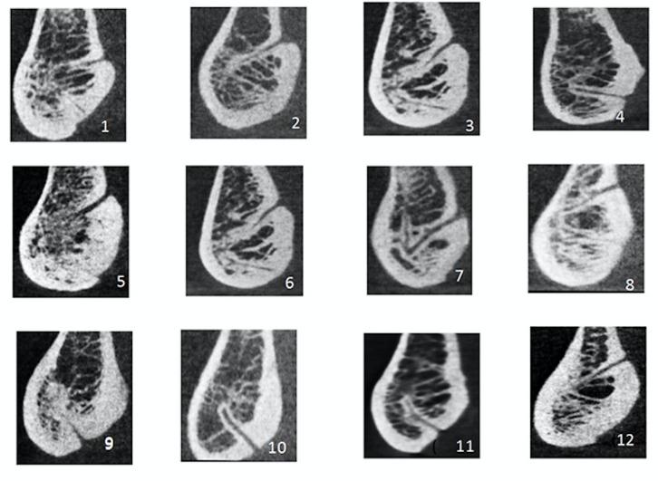
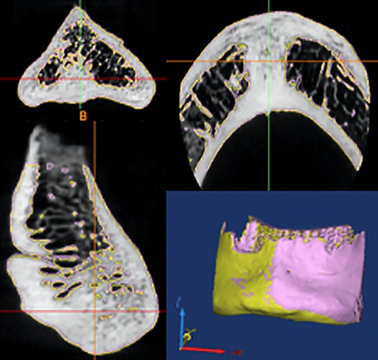
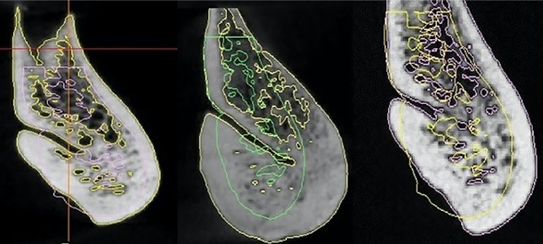
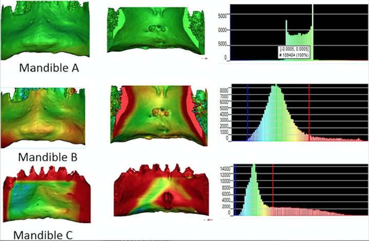
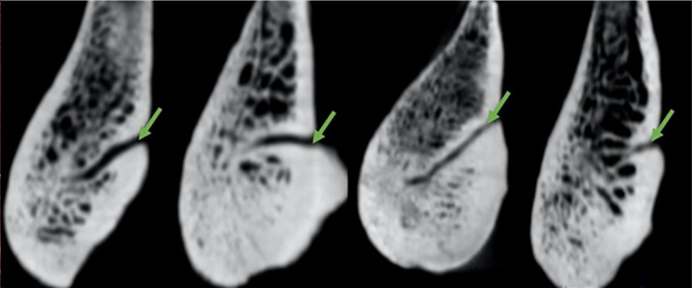
Materials and methods: The study consisted of two parts; an observational part and an objective image analysis part. In the observational part a total of 100 in vivo high resolution CBCT datasets of human mandibles were included in the process of simulated matching of ante-mortem (AM) and post-mortem (PM) data. For the objective image analysis part 10 dry human mandibles were scanned with 2 different Cone Beam Computed tomography (CBCT) machines. In the observational part of the study trained observers attempted to correctly identify matching pairs of images taken from the same mandible out of a series of 100 mandibles. The aim was to simulate matching of the neurovascular structures on AM and PM mandibular midline images and determine the percentage of mandibles identified correctly. In the objective image analysis part, simulated matching was carried out using a specific CBCT dataset acquired to mimic a PM dataset and 10 datasets acquired from a different CBCT device which served as the source of potential AM cases. Comparison between AM and PM datasets resulted in the matching of the AM data and PM data obtained from the same mandible, leading to an assumed correct identification.
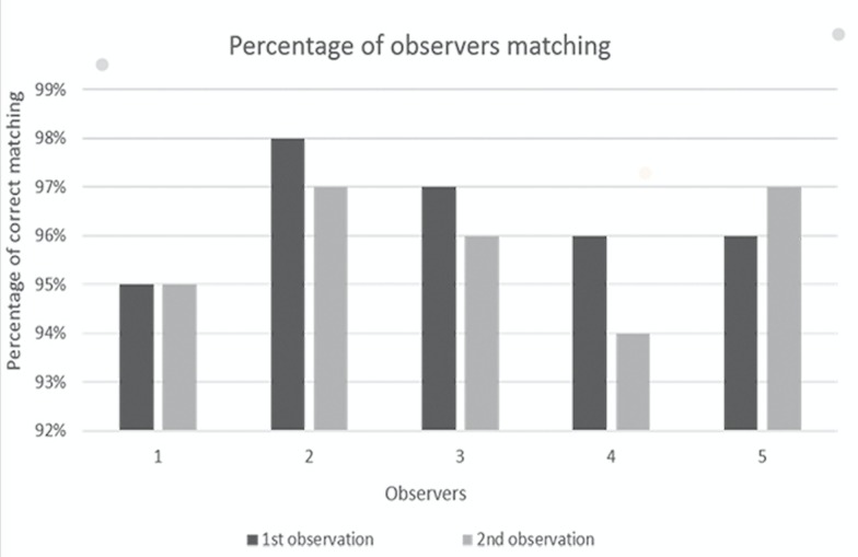
Results: The observational part of the study showed an average 95% correct identification of the mandibular midline neurovascular structures. Registration of mandibles resulted in perfect overlap of the same mandible from 2 different CBCT machine with an error distance equalling zero, while the registration of different mandibles deviated on average error distance 0.13 mm to 0.18 mm.
Conclusion: The percentage of fit for the simulated AM and PM data of the same mandible was 100%. This finding together with the significant deviations noted for the non-matching cases, may have a potential role in forensic identification in the same way that fingerprints are recognised as being a unique identifying feature.
Conflict of interest statement
The authors declare that they have no conflict of interest.
Figures
Similar articles
-
Semi-automatic forensic approach using mandibular midline lingual structures as fingerprint: a pilot study.J Forensic Odontostomatol. 2017 Dec 1;35(2):35-41. J Forensic Odontostomatol. 2017. PMID: 29384735 Free PMC article.
-
Chronologic and geographic variability of neurovascular structures in the human mandible.Forensic Sci Int. 2009 Sep 10;190(1-3):24-32. doi: 10.1016/j.forsciint.2009.05.006. Epub 2009 Jun 13. Forensic Sci Int. 2009. PMID: 19525074
-
Anatomical characteristics of the lingual foramen in ancient skulls: a cone beam computed tomography study in an Anatolian population.Folia Morphol (Warsz). 2018;77(3):514-520. doi: 10.5603/FM.a2018.0009. Epub 2018 Jan 18. Folia Morphol (Warsz). 2018. PMID: 29345723
-
Identification of double mandibular canals: literature review and three case reports with CT scans and cone beam CT.Dentomaxillofac Radiol. 2007 Jan;36(1):34-8. doi: 10.1259/dmfr/27374727. Dentomaxillofac Radiol. 2007. PMID: 17329586 Review.
-
Use of non-clinical smile images for human identification: a systematic review.J Forensic Odontostomatol. 2022 Apr 30;40(1):65-73. J Forensic Odontostomatol. 2022. PMID: 35499538 Free PMC article.
Cited by
-
Cone-Beam Computed Tomography: A New Tool on the Horizon for Forensic Dentistry.Int J Environ Res Public Health. 2022 Apr 28;19(9):5352. doi: 10.3390/ijerph19095352. Int J Environ Res Public Health. 2022. PMID: 35564747 Free PMC article. Review.
-
Semi-automatic forensic approach using mandibular midline lingual structures as fingerprint: a pilot study.J Forensic Odontostomatol. 2017 Dec 1;35(2):35-41. J Forensic Odontostomatol. 2017. PMID: 29384735 Free PMC article.
-
[Use of diagnostic modalities for dentofacial imaging in forensic dentistry. Literature review].Rev Cient Odontol (Lima). 2021 Dec 9;9(4):e088. doi: 10.21142/2523-2754-0904-2021-088. eCollection 2021 Oct-Dec. Rev Cient Odontol (Lima). 2021. PMID: 38463727 Free PMC article. Review. Spanish.
References
-
- Silva RF, Prado MM, Botelho TL, Reges RV, Marinho DEA. Anatomical variations in the permanent mandibular canine: forensic importance. RSBO. 2012;9(4):468–73.
-
- Pillai TJ, Devi TS, Devi CKL. Studies on human mandibles. IOSR Journal of Dental and Medical Science. 2014;13(1):8–15. 10.9790/0853-13120815 - DOI
MeSH terms
LinkOut - more resources
Full Text Sources