

Ultrasound guided double injection of blood into cisterna magna: a rabbit model for treatment of cerebral vasospasm
- PMID: 26851937
- PMCID: PMC4744401
- DOI: 10.1186/s12938-016-0123-z
Ultrasound guided double injection of blood into cisterna magna: a rabbit model for treatment of cerebral vasospasm
Abstract
Background: Double injection of blood into cisterna magna using a rabbit model results in cerebral vasospasm. An unacceptably high mortality rate tends to limit the application of model. Ultrasound guided puncture can provide real-time imaging guidance for operation. The aim of this paper is to establish a safe and effective rabbit model of cerebral vasospasm after subarachnoid hemorrhage with the assistance of ultrasound medical imaging.
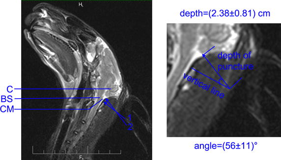
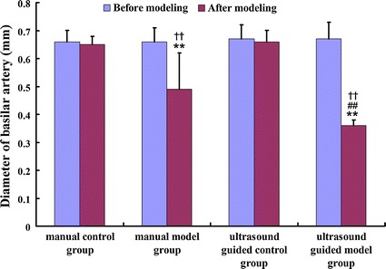
Methods: A total of 160 New Zealand white rabbits were randomly divided into four groups of 40 each: (1) manual control group, (2) manual model group, (3) ultrasound guided control group, and (4) ultrasound guided model group. The subarachnoid hemorrhage was intentionally caused by double injection of blood into their cisterna magna. Then, basilar artery diameters were measured using magnetic resonance angiography before modeling and 5 days after modeling.
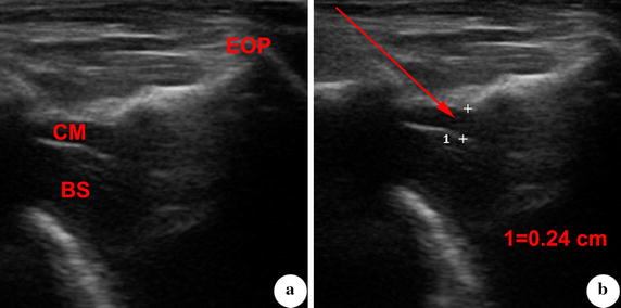
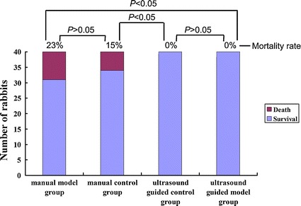
Results: The depth of needle entering into cisterna magna was determined during the process of ultrasound guided puncture. The mortality rates in manual control group and model group were 15 and 23 %, respectively. No rabbits were sacrificed in those two ultrasound guided groups. We found that the mortality rate in ultrasound guided groups decreased significantly compared to manual groups. Compared with diameters before modeling, the basilar artery diameters after modeling were significantly lower in manual and ultrasound guided model groups. The vasospasm aggravated and the proportion of severe vasospasms was greater in ultrasound guided model group than that of manual group. In manual model group, no vasospasm was found in 8 % of rabbits.
Conclusions: The ultrasound guided double injection of blood into cisterna magna is a safe and effective rabbit model for treatment of cerebral vasospasm.
Figures


Similar articles
-
Changes of the Expression and Activity of Phosphodiesterase V in the Basilar Artery Before and After Cerebral Vasospasm in a Rabbit Model.World Neurosurg. 2019 Dec;132:e795-e801. doi: 10.1016/j.wneu.2019.08.008. Epub 2019 Aug 9. World Neurosurg. 2019. PMID: 31404697
-
The single and double blood injection rabbit subarachnoid hemorrhage model.Transl Stroke Res. 2015 Feb;6(1):88-97. doi: 10.1007/s12975-014-0375-5. Epub 2014 Nov 8. Transl Stroke Res. 2015. PMID: 25381219 Review.
-
Effect of cervical sympathetic block on cerebral vasospasm after subarachnoid hemorrhage in rabbits.Acta Cir Bras. 2013 Feb;28(2):89-93. doi: 10.1590/s0102-86502013000200001. Acta Cir Bras. 2013. PMID: 23370920
-
Comparison between one- and two-hemorrhage models of cerebral vasospasm in rabbits.J Neurosci Methods. 2007 Jan 30;159(2):318-24. doi: 10.1016/j.jneumeth.2006.07.026. Epub 2006 Aug 30. J Neurosci Methods. 2007. PMID: 16942802
-
The rabbit blood shunt subarachnoid haemorrhage model.Acta Neurochir Suppl. 2015;120:337-42. doi: 10.1007/978-3-319-04981-6_58. Acta Neurochir Suppl. 2015. PMID: 25366648 Review.
Cited by
-
Magnetic resonance analysis of deep cerebral venous vasospasm after subarachnoid hemorrhage in rabbits.Front Cardiovasc Med. 2022 Sep 21;9:1013610. doi: 10.3389/fcvm.2022.1013610. eCollection 2022. Front Cardiovasc Med. 2022. PMID: 36211577 Free PMC article.
-
Longitudinal imaging and evaluation of SAH-associated cerebral large artery vasospasm in mice using micro-CT and angiography.J Cereb Blood Flow Metab. 2020 Nov;40(11):2265-2277. doi: 10.1177/0271678X19887052. Epub 2019 Nov 21. J Cereb Blood Flow Metab. 2020. PMID: 31752586 Free PMC article.
References
-
- Bar B, MacKenzie L, Hurst RW, Grant R, Weigele J, Bhalla PK, et al. Hyperacute vasospasm after aneurysmal subarachnoid hemorrhage. Neurocrit Care. 2015 - PubMed
Publication types
MeSH terms
LinkOut - more resources
Full Text Sources
Other Literature Sources
Medical
