. 2016 Feb 6:9:8.
doi: 10.1186/s13045-015-0231-4.

Cancer-associated fibroblasts promote the progression of endometrial cancer via the SDF-1/CXCR4 axis

Affiliations

Cancer-associated fibroblasts promote the progression of endometrial cancer via the SDF-1/CXCR4 axis

Fei Teng et al. J Hematol Oncol. .

Abstract

Background: Cancer-associated fibroblasts (CAFs) are believed to play an essential role in cancer initiation and development. However, little research has been undertaken to evaluate the role of CAFs in endometrial cancer (EC) progression. We aim to detect the functional contributions of CAFs to promote progression of EC.

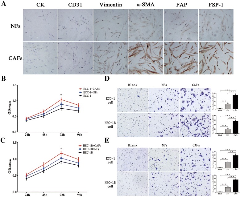
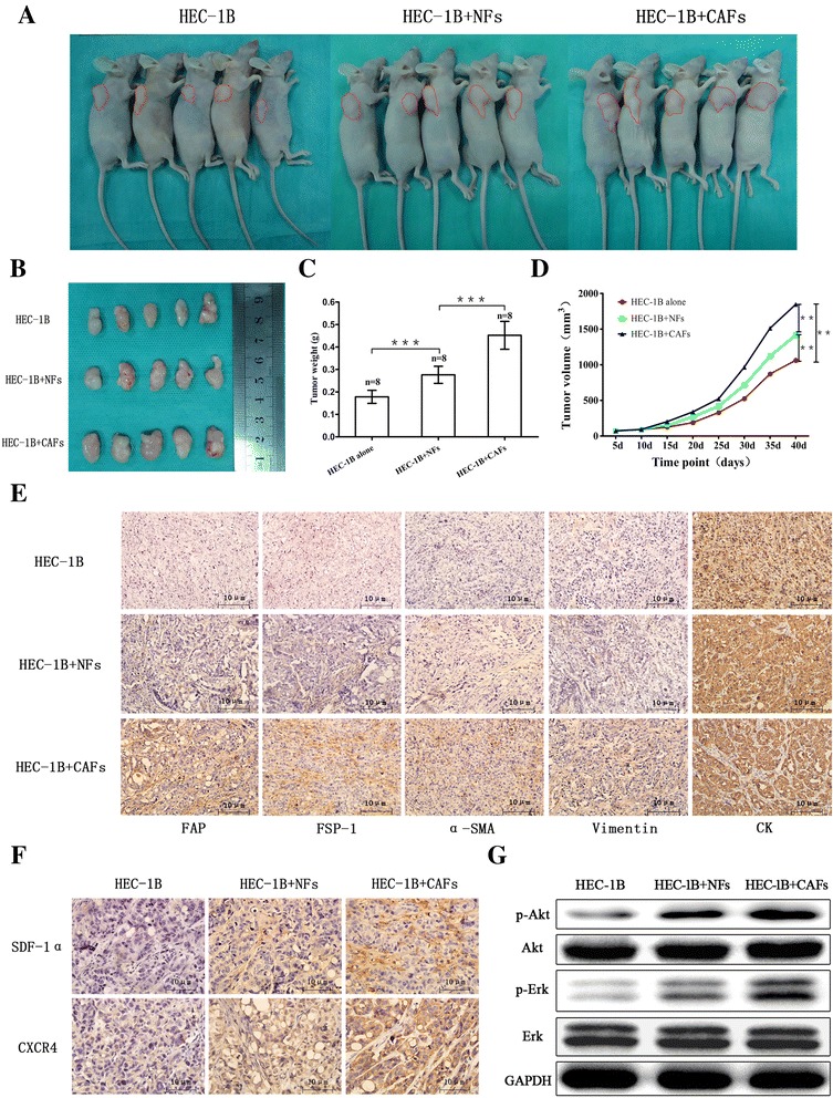
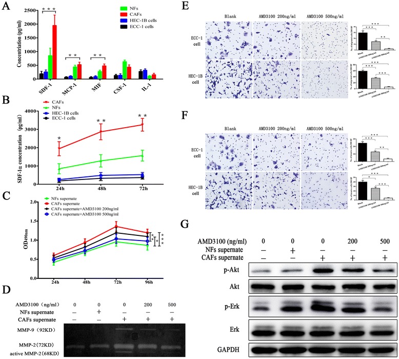
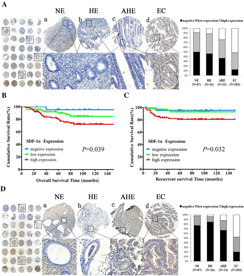
Methods: Stromal fibroblasts were isolated from endometrioid adenocarcinomas and normal endometrial tissues. The conditioned media of cultured CAFs and normal fibroblasts (NFs) were collected to detect the level of stromal cell-derived factor-1alpha (SDF-1α), macrophage chemoattractant protein-1 (MCP-1), migration inhibitory factor (MIF), colony stimulating factor-1 (CSF-1), and interleukin-1 (IL-1) by ELISA. The CAFs or NFs were cocultured with EC cell lines to determine the proliferation, migration, and invasion by MTT assays and transwell chambers. Xenograft models were used to observe tumor growth. Matrix metalloproteinases (MMP)-2 and MMP-9 activity was evaluated by zymography. AMD3100 (a chemokine receptor 4 (CXCR4) antagonist) was used to block the SDF-1/CXCR4 axis. Neutralizing antibodies were used to detect PI3K/Akt and MAPK/Erk pathways by western blotting. SDF-1α and CXCR4 expressions were analyzed in xenotransplanted tumors and 348 cases by immunohistochemistry.

Results: CAFs promoted proliferation, migration, and invasion as well as in vivo tumorigenesis of admixed EC cells significantly more than NFs by secreting SDF-1α. These effects were significantly inhibited by AMD3100. CAFs promoted EC progression via the SDF-1α/CXCR4 axis to activate the PI3K/Akt and MAPK/Erk signalings in a paracrine-dependent manner or increase MMP-2 and MMP-9 secretion in an autocrine-dependent manner. SDF-1α and CXCR4 expression upregulation accompanied clinical EC development and progression. High SDF-1α expression levels were associated with deep myometrial invasion, lymph node metastasis, and poor prognosis in EC.

Conclusions: Our data indicated that CAFs derived from EC tissues promoted EC progression via the SDF-1/CXCR4 axis in a paracrine- or autocrine-dependent manner. SDF-1α is a novel independent poor prognostic factor for EC patients' survival. Targeting the SDF-1/CXCR4 axis might provide a novel therapeutic strategy for EC treatment.


Figures

Fig. 1
Fig. 1
Identification of stromal fibroblasts and CAFs promoting EC cell proliferation, migration, and invasion. a Identification of primary cultured fibroblast cells by immunocytochemistry (magnification ×400). b, c Cell proliferation was measured by MTT assay. d, e Crystal violet staining for migrated and invaded EC cells (magnification ×200). Three independent experiments were performed in triplicate. Data are expressed as means ± SDs. *P < 0.05; **P < 0.01; ***P < 0.001
Fig. 2
Fig. 2
CAFs comingled with HEC-1B cells, enhancing xenograft growth in nude mice. a, b Gross appearance of xenografts; HEC-1B cells were injected alone or were coinjected with various fibroblasts subcutaneously into the right flanks of 4-week-old nude mice, and xenografts were harvested after 40 days of growth in female hosts. c Average tumor weight in each group was evaluated at 40 days after injection. Error bars depict the standard error around the mean. ***P < 0.001. d Tumor volume was plotted on the indicated days. **P < 0.01; ***P < 0.001. e Sections of xenografts in different groups were submitted to immunohistochemistry using antibodies specific for human CK, vimentin, α-SMA, FAP, and FSP-1. Scale bar, 10 μm. f Expression of SDF-1α and CXCR4 in xenotransplanted tumors. g Western blot analysis detection of the phosphorylation of Ark and Erk protein expression in xenotransplanted tumors
Fig. 3
Fig. 3
CAFs promoted EC progression via the SDF-1α/CXCR4 axis. a Cytokines secreted by EC cells, NFs, and CAFs were measured by ELISA. b SDF-1α protein concentration in the conditioned media was measured for 96 h by ELISA. c The effects of SDF-1α-induced proliferation of HEC-1B cells were inhibited by AMD3100. Three independent experiments were performed in triplicate. Data are expressed as means ± SDs. *P < 0.05; **P < 0.01; ***P < 0.001. d MMP-2 and MMP-9 gelatinase activity in the conditioned media was analyzed by zymography. e, f The effects of SDF-1α-induced migration and invasion of EC cells were inhibited by AMD3100 (magnification ×200). g Western blot analysis detection of the phosphorylation of Ark and Erk protein expressions in HEC-1B cells
Fig. 4
Fig. 4
Expression of SDF-1α and CXCR4 in EC. a SDF-1α protein expression upregulation accompanied clinical EC development and progression. b Kaplan-Meier survival curves for overall survival according to SDF-1α staining level: negative expression, low expression, and high expression. Overall survival was significantly different in the three groups (P = 0.039). c Kaplan-Meier survival curves for recurrence according to SDF-1α staining level: negative expression, low expression, and high expression. Recurrent survival was significantly different in the three groups (P = 0.032). d CXCR4 expression increased gradually in endometrial tissues as they progressed from NE to HE to AHE and finally to EC tissues

Similar articles

Cited by

References

    1. Siegel R, Ma J, Zou Z, Jemal A. Cancer statistics. CA Cancer J Clin. 2014;64(1):9–29. doi: 10.3322/caac.21208. - DOI - PubMed
    1. von Gruenigen VE, Waggoner SE, Frasure HE, Kavanagh MB, Janata JW, Rose PG, et al. Lifestyle challenges in endometrial cancer survivorship. Obstet Gynecol. 2011;117(1):93–100. doi: 10.1097/AOG.0b013e31820205b3. - DOI - PubMed
    1. Orimo A, Tomioka Y, Shimizu Y, Sato M, Oigawa S, Kamata K, et al. Cancer-associated myofibroblasts possess various factors to promote endometrial tumor progression. Clin Cancer Res. 2001;7(10):3097–105. - PubMed
    1. Kalluri R, Zeisberg M. Fibroblasts in cancer. Nat Rev Cancer. 2006;6(5):392–401. doi: 10.1038/nrc1877. - DOI - PubMed
    1. Olumi AF, Grossfeld GD, Hayward SW, Carroll PR, Tlsty TD, Cunha GR. Carcinoma-associated fibroblasts direct tumor progression of initiated human prostatic epithelium. Cancer Res. 1999;59(19):5002–11. - PMC - PubMed

Publication types

MeSH terms