Review
. 2016 May;73(9):1765-86.
doi: 10.1007/s00018-016-2147-8. Epub 2016 Feb 6.

Inflammation and its role in age-related macular degeneration

Affiliations
Review

Inflammation and its role in age-related macular degeneration

Anu Kauppinen et al. Cell Mol Life Sci. 2016 May.

Abstract

Inflammation is a cellular response to factors that challenge the homeostasis of cells and tissues. Cell-associated and soluble pattern-recognition receptors, e.g. Toll-like receptors, inflammasome receptors, and complement components initiate complex cellular cascades by recognizing or sensing different pathogen and damage-associated molecular patterns, respectively. Cytokines and chemokines represent alarm messages for leukocytes and once activated, these cells travel long distances to targeted inflamed tissues. Although it is a crucial survival mechanism, prolonged inflammation is detrimental and participates in numerous chronic age-related diseases. This article will review the onset of inflammation and link its functions to the pathogenesis of age-related macular degeneration (AMD), which is the leading cause of severe vision loss in aged individuals in the developed countries. In this progressive disease, degeneration of the retinal pigment epithelium (RPE) results in the death of photoreceptors, leading to a loss of central vision. The RPE is prone to oxidative stress, a factor that together with deteriorating functionality, e.g. decreased intracellular recycling and degradation due to attenuated heterophagy/autophagy, induces inflammation. In the early phases, accumulation of intracellular lipofuscin in the RPE and extracellular drusen between RPE cells and Bruch's membrane can be clinically detected. Subsequently, in dry (atrophic) AMD there is geographic atrophy with discrete areas of RPE loss whereas in the wet (exudative) form there is neovascularization penetrating from the choroid to retinal layers. Elevations in levels of local and systemic biomarkers indicate that chronic inflammation is involved in the pathogenesis of both disease forms.

Keywords: Aging; Eye; Immune system; NLRP3; Retina; Signaling.


Conflict of interest statement

The authors declare that they have no competing interests.

Figures
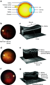
Fig. 1
Fig. 1
a A schematic transverse section through the human eyeball. The macula is located in the posterior pole of the eye. In the center of the macula, a shallow depression in the retina (the fovea) marks the area with the highest visual acuity. Light enters the eye and bends to the sensory retina in the fovea by passing through the transparent media including cornea, lens, and the vitreous body. The sensory retina converts light into nerve impulses, processes the information, and sends it along the visual pathway to the visual cortex. b A normal human retina. A colored photograph of the fundus from the left eye of a healthy subject. The macula is located in the center of the retina. c A cross-section of the normal macula. An OCT scan through the fovea of the healthy left eye reveals the normal organization of the retinal layers. Normal anatomy of the fovea is important for accurate central vision. Modern OCT is an important in vivo tool for ophthalmologists since it allows them to monitor different pathologies non-invasively in this important but tiny and cell-dense location. d A fundus photograph from the left eye of an individual with dry AMD. This demonstrates the presence of numerous yellow deposits, known as drusen, in the central macula. e A cross-section of the macula from an individual with dry AMD. The OCT scan through the fovea of the left eye shows three drusen under the RPE layer. This eye would be expected to suffer from image distortion, as central drusen are prone to reshape the normal foveal pit. Large drusen are associated with decreased visual acuity and disruption of energy homeostasis in the retina. f A fundus photograph from the right eye of an individual with wet AMD. Significant macular edema and exudates together with foveal hemorrhage occur but only small sparse drusen are present centrally. g A cross-section from the macula in the right eye of an individual with wet AMD. An OCT scan through the location of the fovea shows the formation of intraretinal fluid cysts in the fovea. Edema causes the foveal pit to disappear. The local retinal swelling in wet AMD is due to the leaky, abnormal vessels sprouting from the underlying choroid. Intraretinal edema disrupts the normal retinal layer organization and leads to a retinal dysfunction. The OCT scan reveals also a potential hemorrhage and fibrotic lesion development in the fovea. This is another typical finding in wet AMD, likely to result in a permanent central visual field loss, if left untreated. AMD age-related macular degeneration, OCT optical coherence tomography, RPE retinal pigment epithelium
Fig. 2
Fig. 2
Major aspects of TLR signaling. Ligand recognition by LRR domains triggers the dimerisation of TLR proteins. MyD88 and TRIF are the principal adaptor proteins interacting with intracellular TIR domains and mediating the activation of transcription factors, such as NF-κB and IRFs for the production of pro-inflammatory cytokines and type I interferons. IRFs interferon regulatory factors, LRR leucine-rich repeat, MyD88 myeloid differentiation-primary response gene 88, NF-κB nuclear factor kappa B, TIR Toll/IL-1 receptor domain, TRIF TIR-domain-containing adaptor inducing IFN-β
Fig. 3
Fig. 3
Pro-inflammatory inflammasomes. Four NLRs, two DNA sensors, and an RLR are currently the most well-known inflammasomes that promote inflammation by resulting in the release of inflammatory mediators. Receptors lacking the CARD domain are dependent on the adaptor protein ASC for their interaction with pro-caspase 1, which becomes activated by autocleavage into 20 and 10 kDa subunits by the complex assembly
Fig. 4
Fig. 4
Overview of the NLRP3 inflammasome. Ligand recognition through LRR domains results in a conformational change and oligomerization of NLRP3 receptor proteins (a). Thereafter, the adaptor protein ASC binds NLRP3 by PYD–PYD interactions (b). Binding of pro-caspase-1 to ASC through CASP–CASP interactions promotes autocleavage and thereby activation of the caspase-1 enzyme (c). Finally, caspase-1 cleaves the pro-inflammatory cytokines IL-1β and IL-18 into their mature and secreted forms (d)
Fig. 5
Fig. 5
Initiation of the inflammatory response. Recognition of PAMPs and DAMPs by PRRs triggers intracellular signaling resulting in the production of pro-inflammatory cytokines and chemokines. The released mediators contribute to the activation of endothelium, e.g. elevated expression of adhesion molecules and increased vascular permeabilization. Circulating leukocytes interact with adhesion molecules expressed by endothelium, slow down their speed and start rolling along the endothelial layer. The chemokine gradient which originates from the inflamed tissue becomes sensed by leukocytes that start expressing integrins to permit their tighter binding to endothelial cells. Finally, leukocytes leave the circulation to seek out the inflamed tissue where monocytes differentiate into macrophages and dendritic cells according to the local conditions

Similar articles

Cited by

References

    1. Thomas CJ, Schroder K. Pattern recognition receptor function in neutrophils. Trends Immunol. 2013;34:317–328. doi: 10.1016/j.it.2013.02.008. - DOI - PubMed
    1. Lambrecht BN, Hammad H. The airway epithelium in asthma. Nat Med. 2012;18:684–692. doi: 10.1038/nm.2737. - DOI - PubMed
    1. Franceschi C, Bonafe M, Valensin S, Olivieri F, De Luca M, Ottaviani E, De Benedictis G. Inflamm-aging. An evolutionary perspective on immunosenescence. Ann N Y Acad Sci. 2000;908:244–254. doi: 10.1111/j.1749-6632.2000.tb06651.x. - DOI - PubMed
    1. Salvioli S, Capri M, Valensin S, Tieri P, Monti D, Ottaviani E, Franceschi C. Inflamm-aging, cytokines and aging: state of the art, new hypotheses on the role of mitochondria and new perspectives from systems biology. Curr Pharm Des. 2006;12:3161–3171. doi: 10.2174/138161206777947470. - DOI - PubMed
    1. Jarrett SG, Boulton ME. Consequences of oxidative stress in age-related macular degeneration. Mol Aspects Med. 2012;33:399–417. doi: 10.1016/j.mam.2012.03.009. - DOI - PMC - PubMed

Publication types