

Development of a Hybrid Nanoprobe for Triple-Modality MR/SPECT/Optical Fluorescence Imaging
- PMID: 26852675
- PMCID: PMC4665510
- DOI: 10.3390/diagnostics4010013
Development of a Hybrid Nanoprobe for Triple-Modality MR/SPECT/Optical Fluorescence Imaging
Abstract
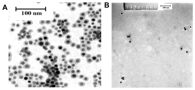
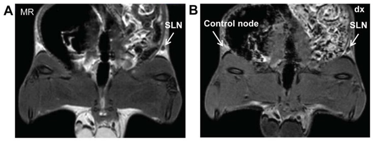
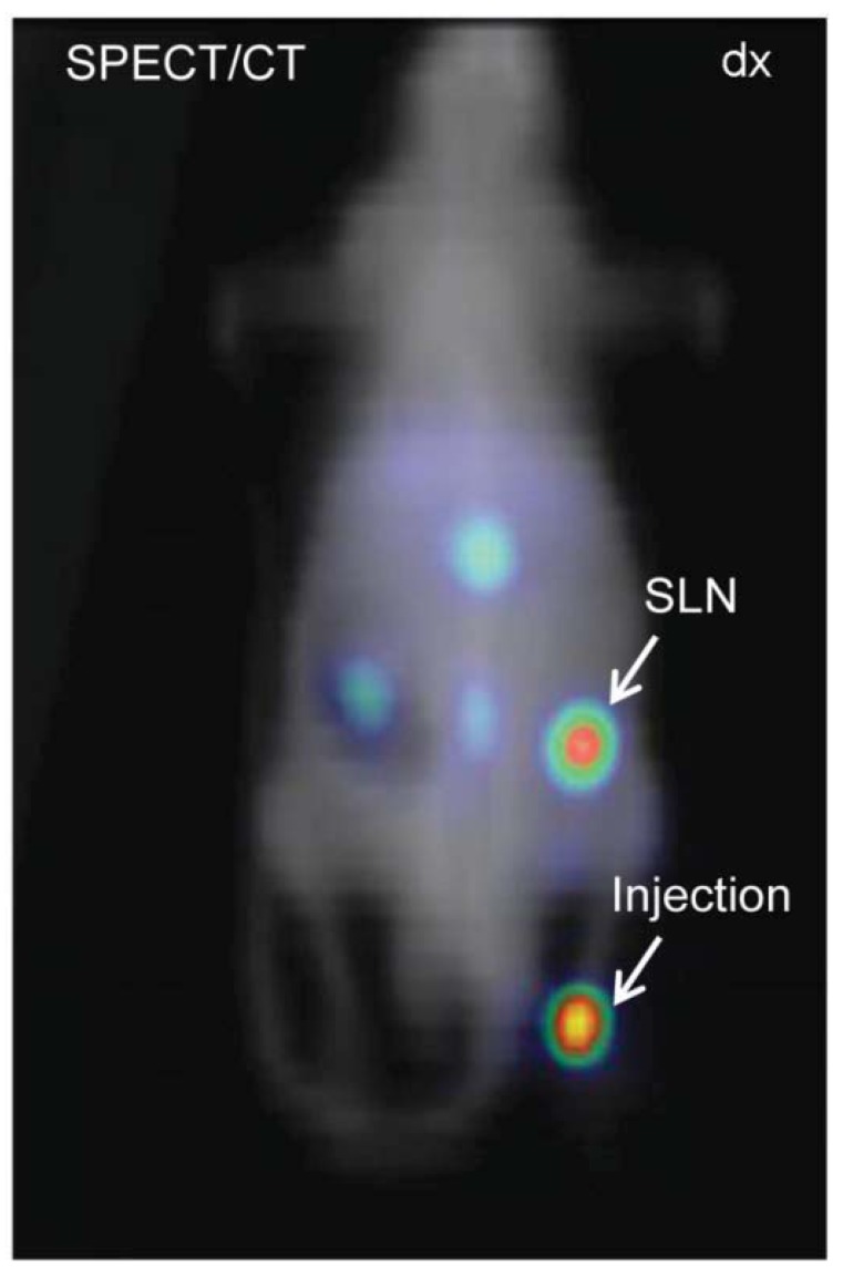
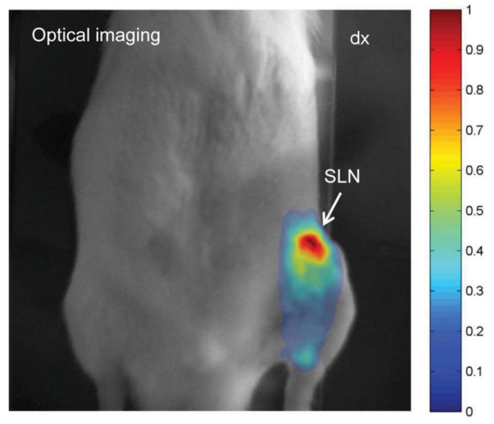
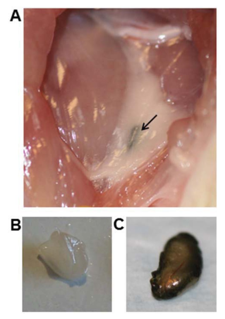
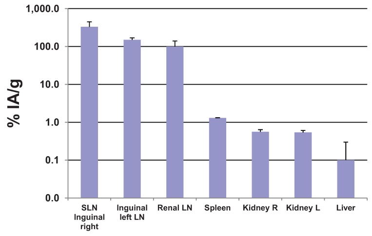
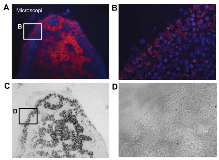
Hybrid clinical imaging is an emerging technology, which improves disease diagnosis by combining already existing technologies. With the combination of high-resolution morphological imaging, i.e., MRI/CT, and high-sensitive molecular detection offered by SPECT/PET/Optical, physicians can detect disease progression at an early stage and design patient-specific treatments. To fully exploit the possibilities of hybrid imaging a hybrid probe compatible with each imaging technology is required. Here, we present a hybrid nanoprobe for triple modality MR/SPECT/Fluorescence imaging. Our imaging agent is comprised of superparamagnetic iron oxide nanoparticles (SPIONs), labeled with (99m)Tc and an Alexa fluorophore (AF), together forming (99m)Tc-AF-SPIONs. The agent was stable in human serum, and, after subcutaneous injection in the hind paw of Wistar rats, showed to be highly specific by accumulating in the sentinel lymph node. All three modalities clearly visualized the imaging agent. Our results show that a single imaging agent can be used for hybrid imaging. The use of a single hybrid contrast agent permits simultaneous hybrid imaging and, more conventionally, allow for single modality imaging at different time points. For example, a hybrid contrast agent enables pre-operative planning, intra-operative guidance, and post-operative evaluation with the same contrast agent.
Keywords: MR; SLN; SPECT; SPION; fluorescence imaging; magnetic resonance imaging; optical.
Figures
Similar articles
-
(68)Ga-labeled superparamagnetic iron oxide nanoparticles (SPIONs) for multi-modality PET/MR/Cherenkov luminescence imaging of sentinel lymph nodes.Am J Nucl Med Mol Imaging. 2013 Dec 15;4(1):60-9. eCollection 2013. Am J Nucl Med Mol Imaging. 2013. PMID: 24380046 Free PMC article.
-
99mTc-labeled superparamagnetic iron oxide nanoparticles for multimodality SPECT/MRI of sentinel lymph nodes.J Nucl Med. 2012 Mar;53(3):459-63. doi: 10.2967/jnumed.111.092437. Epub 2012 Feb 9. J Nucl Med. 2012. PMID: 22323777
-
Simultaneous Preclinical Positron Emission Tomography-Magnetic Resonance Imaging Study of Lymphatic Drainage of Chelator-Free 64Cu-Labeled Nanoparticles.Cancer Biother Radiopharm. 2018 Aug;33(6):213-220. doi: 10.1089/cbr.2017.2412. Epub 2018 Jul 23. Cancer Biother Radiopharm. 2018. PMID: 30036073
-
Main applications of hybrid PET-MRI contrast agents: a review.Contrast Media Mol Imaging. 2016 Mar-Apr;11(2):92-8. doi: 10.1002/cmmi.1674. Epub 2015 Dec 3. Contrast Media Mol Imaging. 2016. PMID: 26632007 Review.
-
Nanoprobes for hybrid SPECT/MR molecular imaging.Nanomedicine (Lond). 2012 May;7(5):719-33. doi: 10.2217/nnm.12.32. Nanomedicine (Lond). 2012. PMID: 22630153 Review.
Cited by
-
Recent Advances in Higher-Order, Multimodal, Biomedical Imaging Agents.Small. 2015 Sep 16;11(35):4445-61. doi: 10.1002/smll.201500735. Epub 2015 Jul 16. Small. 2015. PMID: 26185099 Free PMC article. Review.
References
LinkOut - more resources
Full Text Sources
Other Literature Sources
