. 2016 Feb 18;61(4):602-613.
doi: 10.1016/j.molcel.2016.01.009. Epub 2016 Feb 4.

Structural Basis and Functional Role of Intramembrane Trimerization of the Fas/CD95 Death Receptor

Affiliations

Structural Basis and Functional Role of Intramembrane Trimerization of the Fas/CD95 Death Receptor

Qingshan Fu et al. Mol Cell. .

Abstract

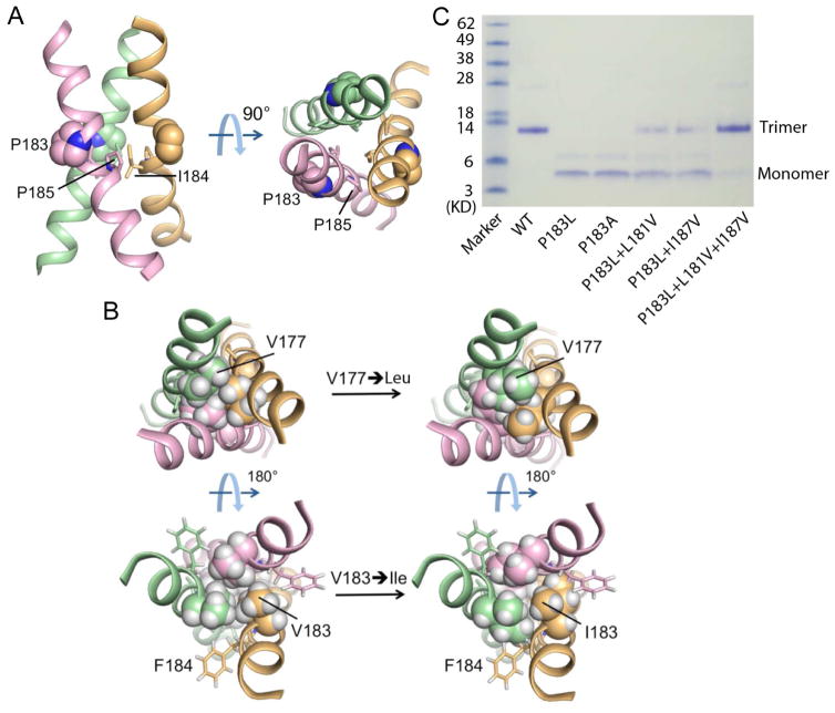
Fas (CD95, Apo-1, or TNFRSF6) is a prototypical apoptosis-inducing death receptor in the tumor necrosis factor receptor (TNFR) superfamily. While the extracellular domains of TNFRs form trimeric complexes with their ligands and the intracellular domains engage in higher-order oligomerization, the role of the transmembrane (TM) domains is unknown. We determined the NMR structures of mouse and human Fas TM domains in bicelles that mimic lipid bilayers. Surprisingly, these domains use proline motifs to create optimal packing in homotrimer assembly distinct from classical trimeric coiled-coils in solution. Cancer-associated and structure-based mutations in Fas TM disrupt trimerization in vitro and reduce apoptosis induction in vivo, indicating the essential role of intramembrane trimerization in receptor activity. Our data suggest that the structures represent the signaling-active conformation of Fas TM, which appears to be different from the pre-ligand conformation. Analysis of other TNFR sequences suggests proline-containing sequences as common motifs for receptor TM trimerization.


Figures

Figure 1
Figure 1. NMR and Biochemical Characterization of the TM Domains of Mouse and Human Fas Receptors
(A) Sequence alignment between the mouse and human Fas TM regions. The alignment is based on ClustalW2. (mFAS: mouse FAS; hFAS: human FAS) (B and C) SDS-PAGE analysis of the TM domain oligomerization in NMR sample condition for mouse (B) and human (C) Fas receptors. The gel lanes from left to right are: (1) purified peptide powder dissolved in gel loading buffer; (2) purified peptide reconstituted in DMPC/DHPC bicelles (q = 0.5) as in the preparation of the NMR samples; (3) M.W. standards. Samples were run under non-denaturing conditions. (D and E) Two-dimensional 1H-15N TROSY-HSQC spectra of the TM trimers of mouse (D) and human (E) Fas receptors reconstituted in DMPC/DHPC bicelles (q = 0.5) recorded at 600 MHz. The peptides are uniformly (15N, 13C, 2H)-labeled.
Figure 2
Figure 2. Structures of the Trimeric TM Domains of Fas in Bicelles
(A) Ensemble of 15 low energy structures calculated using NMR-derived restraints. The backbone and all heavy atom RMSDs are 0.829 Å and 1.392 Å for mouse (left) and are 0.859 Å and 1.605 Å for human (right). See Table 1 for more statistics. (B) Ribbon representations of the mouse Fas-TM trimers showing residues that form the hydrophobic core. (C) Ribbon representations of the human Fas-TM trimers. (D) Superposition of human FAS-TM trimer (yellow) and canonical coiled-coil trimer (blue, PDB: 2NVL). (E) Helical wheel illustration of standard coiled coil trimer showing 3.5 residues per helical turn and core positions ‘a’ and ‘d’ shaded in green. The positions ‘g’ highlighted in yellow are periphery in standard coiled coil. (F) Helical wheel illustration of Fas-TM trimer showing three positions, ‘a’, ‘d’, and ‘g’, involved in forming the core. (G) Zoomed side view showing inter-helical VDW interactions between the central proline at position i and isoleucine at position i-1.
Figure 3
Figure 3. Effects of Single Mutations on Trimerization of the Fas TM Domains
(A) Helical wheel illustration of mouse Fas-TM trimer as in Figure 2E showing the positions of single mutation highlighted in green. The dashed green lines indicate VDW contact between two residue positions. (B) SDS-PAGE of bicelle-reconstituted mouse Fas-TM and its mutants showing the effect of single mutations on trimerization. Samples were run under non-denaturing conditions. (C) Helical wheel representation as in (A) for the human Fas-TM trimer showing the positions of single mutation in green as well as the disease mutation positions in yellow. (D) SDS-PAGE of bicelle-reconstituted human Fas-TM and its mutants showing the effect of single mutations on trimerization. Samples were run under non-denaturing conditions.
Figure 4
Figure 4. Role of the Lipid-Facing Proline in the Trimeric Assembly of Human Fas-TM
(A) Ribbon representations of human Fas-TM trimer showing the lipid-facing proline (P183) as spheres. The central proline (P185) is shown as sticks. (B) Top and bottom views of mouse Fas-TM trimer showing that mutating V177 or V183 to isoleucine results in severe steric collisions on either side of the central proline. (C) SDS-PAGE of bicelle reconstituted human Fas-TM and its mutants showing the effect of single mutations at P183 as well as compensatory mutations on trimerization.
Figure 5
Figure 5. Trimerization of the TM Domain is Essential for Overexpression-Induced Fas Killing
(A–J) HeLa cells transiently overexpressed with WT and mutant Fas were stained by PI and annexin-V, and analyzed by Cellometer. One representative data set out of three were shown. High PI and/or high annexin-V stained cells were considered dead cells. (K) Plotted cell death ratios in WT and mutant Fas-overexpressed HeLa cells. The data showed that C178R, P183L and P185A mutant Fas were partially defective in cell killing compared to WT Fas. Error bars were derived from three independent experiments.
Figure 6
Figure 6. Effects of Fas-TM Domain Mutants on Receptor Pre-Association and FasL-Induced Apoptosis
(A, B) Relative FRET between the indicated CFP and YFP fusion proteins of Fas in transfected 293T cells. The fold change in FRET relative to WT-WT interactions for heterotypic mutant:WT (A) and homotypic (B) interactions are shown. Data are from two independent experiments with each repeat shown as one point, and are representative of five independent experiments. Statistical comparisons to WTFas-TNFR1 association (A) and WTFas self-association (B) are shown (*p ≤ 0.05; **p ≤ 0.01; ***p ≤ 0.001 by Mann-Whitney). (C) Oligomerization state of WT, P183L and L180F Fas mutants on the plasma membrane of COS-7 assessed by quantitative analysis of PALM data sets. The spatial distribution of single PAGFP-tagged CD95, CD95-P183L and CD95-L180F across the plasma membrane were analyzed by Hoshen-Kopelman based algorithm to identify individual protein clusters and the number of proteins within each identified cluster was counted. The plot shows the frequency of occurrence of clusters containing specified number of proteins, and the percentage of clustered molecules are shown in the legend. (D) Fas-deficient Jurkat RapoC2 cells were transfected with either WT Fas or the indicated TM mutation and stimulated with increasing amounts of FasL-LZ for 18 hours to induce apoptosis. Data shown are cumulative of 3 independent experiments each performed in triplicate represented as mean +/- SEM. Unpaired t-test was used for statistical analyses. **p ≤ 0.01, ****p ≤ 0.0001 for all concentrations above 2.5 ng ml−1 of the indicated mutant compared to WT. (E) Dominant negative interference assay with Death-domain-truncated (ΔDD) Fas-YFP fusion constructs co-transfected with full-length Fas-CFP fusion constructs into Jurkat RapoC2 cells. Transfected cells were stimulated with 25 ng/ml FasL-LZ for 18hr and CFP+YFP+ cells of equivalent relative fluorescence were assessed for cell death. Asterisks represent significance (p < 0.005) for comparison of the ability of mutant Fas constructs to confer resistance to FasL-induced apoptosis compared with the WT ΔDD protein. Data are cumulative of two independent experiments, each performed in triplicate.
Figure 7
Figure 7. Implication of Fas-TM Trimerization to Transmembrane Signaling By TNFRs
(A) TM domain sequences within different receptor families of the TNFR superfamily, aligned using the φPxφ motif. (B) Schematic illustration of the role of proline motif mediated Fas-TM trimerization in stabilizing the trimeric receptor conformation that is signaling active. Dimeric pre-ligand association is supported by this study and observations of crystallographic dimers in the unliganded ECD of TNFR1 (Naismith et al., 1995). Higher-order clustering of liganded Fas is supported by structural and functional studies of intracellular interactions.

References

    1. Arkhipov A, Shan Y, Das R, Endres NF, Eastwood MP, Wemmer DE, Kuriyan J, Shaw DE. Architecture and membrane interactions of the EGF receptor. Cell. 2013;152:557–569. - PMC - PubMed
    1. Call ME, Pyrdol J, Wiedmann M, Wucherpfennig KW. The organizing principle in the formation of the T cell receptor-CD3 complex. Cell. 2002;111:967–979. - PMC - PubMed
    1. Call ME, Schnell JR, Xu C, Lutz RA, Chou JJ, Wucherpfennig KW. The structure of the zetazeta transmembrane dimer reveals features essential for its assembly with the T cell receptor. Cell. 2006;127:355–368. - PMC - PubMed
    1. Call ME, Wucherpfennig KW, Chou JJ. The structural basis for intramembrane assembly of an activating immunoreceptor complex. Nature immunology. 2010;11:1023–1029. - PMC - PubMed
    1. Chan FK. The pre-ligand binding assembly domain: a potential target of inhibition of tumour necrosis factor receptor function. Ann Rheum Dis. 2000;59(Suppl 1):i50–53. - PMC - PubMed

Publication types