. 2016 Feb 8:7:10596.
doi: 10.1038/ncomms10596.

Mechanical evidence that Australopithecus sediba was limited in its ability to eat hard foods

Affiliations

Mechanical evidence that Australopithecus sediba was limited in its ability to eat hard foods

Justin A Ledogar et al. Nat Commun. .

Abstract

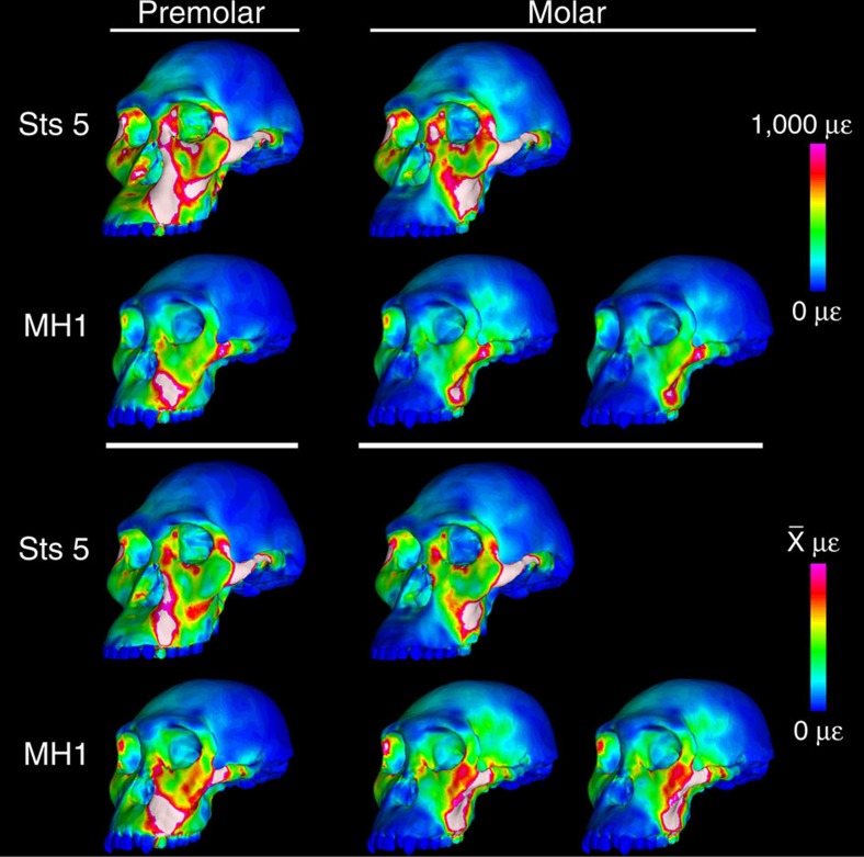
Australopithecus sediba has been hypothesized to be a close relative of the genus Homo. Here we show that MH1, the type specimen of A. sediba, was not optimized to produce high molar bite force and appears to have been limited in its ability to consume foods that were mechanically challenging to eat. Dental microwear data have previously been interpreted as indicating that A. sediba consumed hard foods, so our findings illustrate that mechanical data are essential if one aims to reconstruct a relatively complete picture of feeding adaptations in extinct hominins. An implication of our study is that the key to understanding the origin of Homo lies in understanding how environmental changes disrupted gracile australopith niches. Resulting selection pressures led to changes in diet and dietary adaption that set the stage for the emergence of our genus.


Figures

Figure 1
Figure 1. Line plot of von Mises strain generated during simulated biting in finite element models.
Strain data correspond to (a) left premolar (P3) and (b) left molar biting (M2), recorded from 14 homologous locations across the craniofacial skeleton of finite element models of Sts 5 (A. africanus) and MH1 (A. sediba). The grey region brackets the range of variation exhibited by six chimpanzee crania intentionally selected to be morphologically different. For the molar biting analysis, it was necessary to rerun the model of MH1 with the balancing (non-biting) side muscle forces reduced by nearly 30% to remove a distractive (tensile) reaction force at the working (biting) side jaw joint. Therefore, data for both the symmetrical (S) and asymmetrical (A) loadings are shown.
Figure 2
Figure 2. Colour mapping of von Mises strain in finite element models of Sts 5 (A. africanus) and MH1 (A. sediba) crania during simulated left premolar (P3) and left molar (M2) biting (not to scale).
Colour maps on the top half of the figure reflect absolute strain magnitudes ranging from 0 to 1,000 microstrain (μɛ), where white regions experience strain magnitudes that exceed 1,000 μɛ. Colour maps on the bottom half of the figures reflect relative strain magnitudes where the colour scale of each model ranges from 0 to a value equal to twice the average of strain magnitudes collected from 10 standard locations from which strain data have been collected during in vivo feeding experiments in primates. These relative strain maps provide information about the distribution of relatively high and relatively low strain concentrations independent of the scale of the strain magnitudes. For the molar biting analysis, it was necessary to rerun the model of MH1 with the balancing (non-biting) side muscle forces reduced by nearly 30% in order to remove a distractive (tensile) reaction force at the working (biting) side jaw joint. Therefore, under the column for molar biting, the model of MH1 is shown loaded with bilaterally symmetrical muscle forces (left), as well as with asymmetrical muscle forces (right).
Figure 3
Figure 3. Ontogenetic changes among the dentition, malar root and temporomandibular joint in 319 extant African apes and humans.
(a) Principal component summary of shape differences, represented by mean configurations, among specimens with no molars (open symbols), M2 in occlusion (partially filled symbols) and adults (filled symbols). (b) Surface reconstruction of MH1 specimen rendered from down-sampled synchotron scans (right) and hypothetical adult morphology of MH1 generated using a male chimpanzee developmental trajectory (left).
Figure 4
Figure 4. The constrained lever model of jaw biomechanics.
A ‘triangle of support' is formed by the bite point (BITE) and the working-side (WS) and balancing-side (BS) temporomandibular joints (TMJ). During a premolar bite (a), the muscle resultant vector (MRV) of the jaw adductor (masticatory) muscles remains within the triangle (passing into the plane of the image), producing compression (green circles) at all three points as the mandible is elevated. However, during some molar bites (b), the MRV falls outside the triangle when the muscles are being recruited equally on both sides of the head, producing compression at the bite point and BS TMJ, but distraction (red circle) at the WS TMJ. To eliminate the distraction, the recruitment of the balancing-side muscles must be lessened, thereby causing the MRV to shift its position towards the working side (arrow). Once the MRV falls back within the triangle, then the WS TMJ will be in compression. A consequence of reducing the recruitment of the balancing-side muscles is that the magnitude of the bite force is reduced.
Figure 5
Figure 5. Orientation of the joint reaction force at the working (biting) side temporomandibular joint (TMJ) in models of (MH1) (A. sediba) and Sts 5 (A. africanus) during simulated left premolar (P3) and left molar (M2) biting.
Arrows indicate direction of the reaction force. Yellow arrows indicate a compressive force, while red arrows indicate a distractive force, relative to the plane of the triangle of support (green line). Note that the zygomatic root (off of which the masseter muscle arises) is more mesially positioned relative to the tooth row in MH1 than Sts 5. Models are not shown to scale.

References

    1. Scott R. S., Ungar P. S., Bergstrom T. S. & Brown C. A. Dental microwear texture analysis shows within-species diet variability in fossil hominins. Nature 436, 693–695 (2005). - PubMed
    1. Sponheimer M. et al.. Isotopic evidence for dietary variability in the early hominin Paranthropus robustus. Science 314, 980–982 (2006). - PubMed
    1. Ungar P. S., Grine F. E. & Teaford M. F. Dental microwear and diet of the Plio-Pleistocene hominin Paranthropus boisei. PLOS ONE 3, e2044 (2008). - PMC - PubMed
    1. van der Merwe N. J., Masao F. T. & Bamford M. K. Isotopic evidence for contrasting diets of early hominins Homo habilis and Australopithecus boisei of Tanzania. S. Afr. J. Sci. 104, 153–155 (2008).
    1. Lee-Thorp J. A., Sponheimer M., Passey B. H., de Ruiter D. J. & Cerling T. E. Stable isotopes in fossil hominin tooth enamel suggest a fundamental dietary shift in the Pliocene. Philos. Trans. R. Soc. Lond. B Biol. Sci. 365, 3389–3396 (2010). - PMC - PubMed

Publication types

LinkOut - more resources