. 2016 Feb 8:6:20121.
doi: 10.1038/srep20121.

H19 activates Wnt signaling and promotes osteoblast differentiation by functioning as a competing endogenous RNA

Affiliations

H19 activates Wnt signaling and promotes osteoblast differentiation by functioning as a competing endogenous RNA

Wei-Cheng Liang et al. Sci Rep. .

Abstract

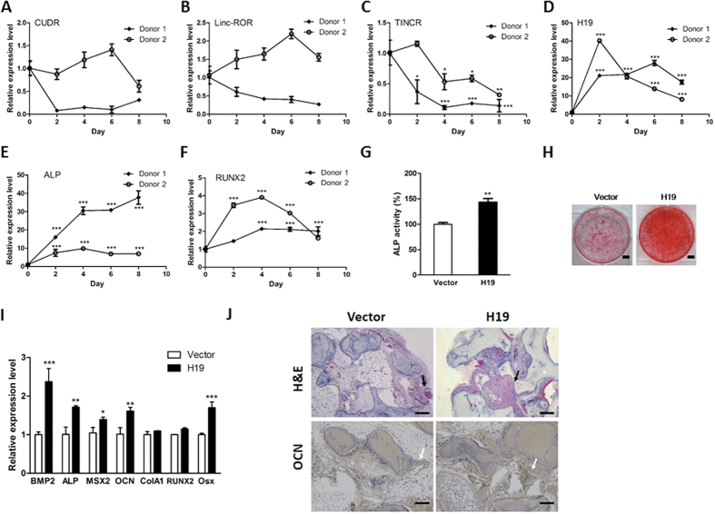
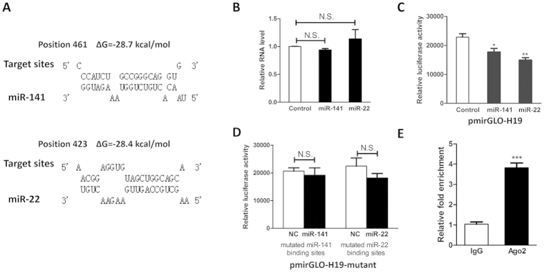
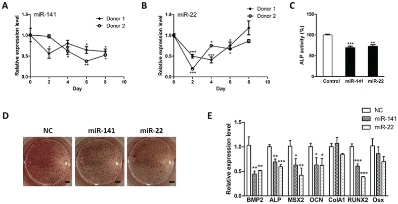
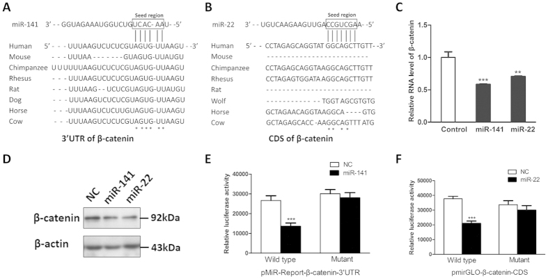
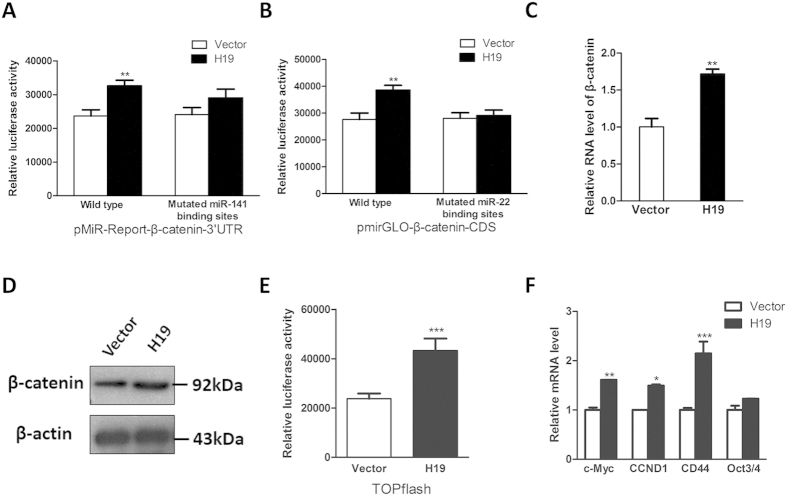
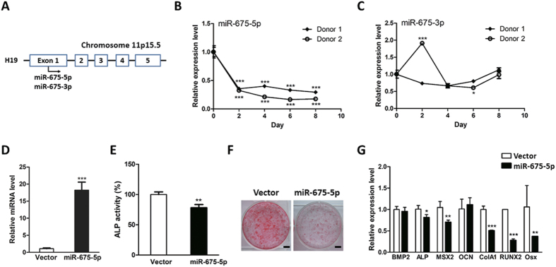
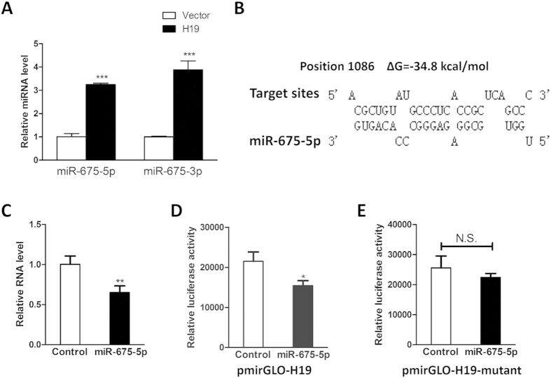
Bone homeostasis is tightly orchestrated and maintained by the balance between osteoblasts and osteoclasts. Recent studies have greatly expanded our understanding of the molecular mechanisms of cellular differentiation. However, the functional roles of non-coding RNAs particularly lncRNAs in remodeling bone architecture remain elusive. In our study, lncRNA H19 was found to be upregulated during osteogenesis in hMSCs. Stable expression of H19 significantly accelerated in vivo and in vitro osteoblast differentiation. Meanwhile, by using bioinformatic investigations and RIP assays combined with luciferase reporter assays, we demonstrated that H19 functioned as an miRNA sponge for miR-141 and miR-22, both of which were negative regulators of osteogenesis and Wnt/β-catenin pathway. Further investigations revealed that H19 antagonized the functions of these two miRNAs and led to de-repression of their shared target gene β-catenin, which eventually activated Wnt/β-catenin pathway and hence potentiated osteogenesis. In addition, we also identified a novel regulatory feedback loop between H19 and its encoded miR-675-5p. And miR-675-5p was found to directly target H19 and counteracted osteoblast differentiation. To sum up, these observations indicate that the lncRNA H19 modulates Wnt/β-catenin pathway by acting as a competing endogenous RNA, which may shed light on the functional role of lncRNAs in coordinating osteogenesis.


Figures

Figure 1
Figure 1. Overexpression of H19 promoted osteogenesis.
(AF) The hMSCs isolated from two independent donors’ bone marrow were initiated by osteogenic medium and harvested at indicated time points. The expression levels of several candidate lncRNAs, including CUDR (A), Linc-ROR (B), TINCR (C), H19 (D), ALP (E) and RUNX2 (F) were monitored by qRT-PCR. (G) The osteogenesis of H19-overexpressing hMSCs was initiated by osteogenic medium. At day 10 post-induction, the ALP activity was measured and ectopic expression of H19 enhanced ALP activity. (H) The osteoblast differentiation of H19-overexpressing hMSCs was started by osteogenic medium. At day 20 post-induction, the calcified nodules were visualized by Alizarin Red stainning and it was found that overexpression of H19 promoted hMSC osteogenesis. Scale bars: 1cm. (I) The RNA levels of osteogenesis-related marker genes were evaluated by qRT-PCR and ectopic expression of H19 upregulated the expression serial osteogenic maker genes. (J) H19-overexpressing hMSCs and corresponding control cells were implanted subcutaneously into the dorsal surfaces of nude mice. At 8 weeks post-implantation, the transplants were collected for histological analysis. Representative images of H&E staining and immunohistochemical staining of OCN were captured. The new bone matrix was indicated by black arrow and OCN proteins were highlighted by white arrow. Scale bars: 100 μm. The results of in vivo bone formation assay showed that H19 significnatly enhanced osteogenesis. (n = 3; *P < 0.05; **P < 0.01; ***P < 0.001).
Figure 2
Figure 2. H19 was a bona fide target for miR-141 and miR-22.
(A) Schematic diagrams of the mutual interplays between miRNA and H19. The positions of miRNA binding sites and calculated ΔG values are showed on the top (kcal/mol). (B) qRT-PCR results showed the relative RNA level of H19 after ectopic expression of miR-141 and miR-22. Neither miR-141 nor miR-22 affected the RNA level of H19. (C) HEK293 cells were transfected with miRNA mimics combined with luciferase reporter harboring H19 gene. The effects of miR-141 and miR-22 on the luciferase activity were measured by luciferase reporter assays. MiR-141 and miR-22 could suppress the luciferase activity. (D) The miR-141 and miR-22 binding sites were mutated and the mutated luciferase reporters were co-transfected with corresponding miRNAs. The mutations on binding sites abolished the previously suppressive effect. (E) H19 RNA level in the immunoprecipitates were measured by qRT-PCR. H19 RNA was enriched in the Ago2-coated beads. (n = 3; *P < 0.05; **P < 0.01; ***P < 0.001).
Figure 3
Figure 3. Overexpression of miR-141 and miR-22 suppressed osteoblast differentiation.
(A,B) The expression levels of miR-141 (A) and miR-22 (B) were determined by qRT-PCR. These two miRNAs were downregulated during osteogenesis. (C) The hMSCs were tranfected with miR-141 or miR-22 mimics. The hMSC osteogenesis was initiated by osteogenic medium. At day 10 post-induction, the ALP activity was measured and ectopic expression of both miR-141 and miR-22 impaired ALP activity. (D) The osteoblast differentiation of miR-141- or miR-22-overexpressing hMSCs was started by osteogenic medium. At day 20 post-induction, the calcified nodules were visualized by Alizarin Red stainning and it was showed that overexpression of miR-141 or miR-22 inhibited hMSC osteogenesis. Scale bars: 1 cm. (E) The RNA levels of osteogenesis-related marker genes were evaluated by qRT-PCR and ectopic expression of miR-141 or miR-22 downregulated the expression serial osteogenic maker genes. (n = 3; *P < 0.05; **P < 0.01; ***P < 0.001).
Figure 4
Figure 4. β-catenin was a bona fide target for miR-141 and miR-22.
(A) The 3′-UTR region of β-catenin harbors a putative miR-141 binding site, which is highly conserved across multiple species. (B) The protein-coding region of β-catenin contains a potential miR-22 binding site, which is conserved across a number of species. (C) Overexpression of miR-141 or miR-22 dramatically reduced the mRNA levels of β-catenin in hMSCs. (D) Overexpression of miR-141 or miR-22 decreased the protein levels of β-catenin in hMSCs. (E) The interaction between miR-141 and 3′UTR region of β-catenin was confirmed by luciferase reporter assays in HEK293 cells. (F) The interaction between miR-22 and protein-coding region of β-catenin was verfied by luciferase reporter assays in HEK293 cells. (n = 3; *P < 0.05; **P < 0.01; ***P < 0.001).
Figure 5
Figure 5. Upregulation of H19 increased the expression of β-catenin and activated Wnt/β-catenin pathway.
(A) Luciferase reporter assays were utilized to monitor the interaction between H19 and luciferase reporters containing the 3′UTR region of β-catenin. Overexpression of H19 upregulated the luciferase activity of wild type luciferase reporter while mutation on miR-141 binding sites counteracted this upregulation effect. (B) Luciferase reporter assays were used to evaluate the interplay between H19 and luciferase reporters containing the partial coding region of β-catenin. Ectopic expression of H19 upregulated the luciferase activity of wild type luciferase reporter while mutation on miR-22 binding sites eliminated this upregulation effect. (C,D) Upregulation of H19 increased the RNA (C) and protein (D) levels of β-catenin. (E) Overexpression of H19 enhanced the TOPflash luciferase activity. (F) Upregulation of H19 activated some downstream transcriptional targets of β-catenin. (n = 3; *P < 0.05; **P < 0.01; ***P < 0.001).
Figure 6
Figure 6. Overexpression of miR-675-5p suppressed osteoblast differentiation.
(A) The schematic representation of the H19 transcript and its encoded miR-675. (B,C) The expression levels of miR-675-5p (B) and miR-675-3p (C) were determined by qRT-PCR. (D) The hMSCs were tranfected with miR-675-5p mimics and the effects of transient transfection were confirmed by qRT-PCR. (E) The osteogenesis of miR-675-5p-overexpressing hMSCs was initiated by osteogenic medium. At day 10 post-induction, the ALP activity was measured and ectopic expression of miR-675-5p impaired ALP activity. (F) The osteoblast differentiation of miR-675-5p-overexpressing hMSCs was started by osteogenic medium. At day 20 post-induction, the calcified nodules were visualized by Alizarin Red stainning and it was showed that overexpression of miR-675-5p suppressed hMSC osteogenesis. Scale bars: 1 cm. (G) The RNA levels of osteogenesis-related marker genes were evaluated by qRT-PCR and ectopic expression of miR-675-5p downregulated the expression serial osteogenic maker genes. (n = 3; *P < 0.05; **P < 0.01; ***P < 0.001).
Figure 7
Figure 7. Overexpression of H19 gave rise to miR-675-5p and miR-675-3p while miR-675-5p downregulated the expression of H19.
(A) The expression levels of miR-675-5p and miR-675-3p were determined by qRT-PCR in the H19-overexpressing hMSCs. Ectopic expression of H19 significantly increased the expression of miR-675-5p and miR-675-3p. (B) Schematic diagrams of the mutual interplays between miR-675-5p and H19. The positions of miR-675-5p binding sites and calculated ΔG values are showed on the top (kcal/mol). (C) qRT-PCR results showed the relative RNA level of H19 after ectopic expression of miR-675-5p. Overexpression of miR-675-5p downregulated the RNA level of H19. (D) HEK293 cells were transfected with miR-675-5p mimics combined with luciferase reporter harboring H19 gene. The effects of miR-675-5p on the luciferase activity were measured by luciferase reporter assays and overexpression of miR-675-5p could suppress the luciferase activity. (E) The miR-675-5p binding sites within luciferase reporter plasmid were mutated. HEK293 cells were transfected with miR-675-5p mimics combined with mutated luciferase reporter harboring H19 gene. The mutatation abolished the suppressive effect. (n = 3; *P < 0.05; **P < 0.01; ***P < 0.001).

Similar articles

Cited by

References

    1. Long F. Building strong bones: molecular regulation of the osteoblast lineage. Nature reviews. Molecular cell biology 13, 27–38, doi: 10.1038/nrm3254 (2012). - DOI - PubMed
    1. Rosenberg N., Rosenberg O. & Soudry M. Osteoblasts in bone physiology-mini review. Rambam Maimonides medical journal 3, e0013, doi: 10.5041/RMMJ.10080 (2012). - DOI - PMC - PubMed
    1. Phan T. C. A., Xu J. & Zheng M. H. Interaction between osteoblast and osteoclast: impact in bone disease. Histol Histopathol 19, 1325–1344 (2004). - PubMed
    1. Pittenger M. F. et al. Multilineage potential of adult human mesenchymal stem cells. Science 284, 143–147 (1999). - PubMed
    1. Zhang W. X., Yang N. L. & Shi X. M. Regulation of mesenchymal stem cell osteogenic differentiation by glucocorticoid-induced leucine zipper (GILZ). J Biol Chem 283, 4723–4729 (2008). - PubMed

Publication types

MeSH terms